Cervical cancer:
Cervical cancer is the third most common gynecologic malignancy
in the United States [4,13]. Almost
half of the cases of invasive cervical cancer occur in patients
under the age of 35 years [13]. About 90% of cervical cancers are
squamous cell, with adenocarcinoma much less common, and cervical sarcoma, lymphoma, and
neuroendocrine tumor being rare [21].
Risk factors for cervical carcinoma include: Multiple sex
partners, condyloma (more than 90% of
squamous cell cancers of the cervix
contain human papilloma virus DNA
[3]), and herpes.
About 85-95% of cervical carcinomas are of the squamous cell variety, and adenocarcinoma accounts for the remaining
5-15%. Predictors of disease free survival in patients with early
stage cervical carcinoma include tumor size, depth of invasion,
and invasion of the lymphatic or vascular space.
Cervical carcinoma most commonly metastasizes to regional lymph nodes and the extent of nodal involvement is an important prognostic indicator [4]. The presence of metstases is directly related to the size of the tumor.
Staging:
FIGO staging of cervical carcinoma is as follows [20,24]:
|
Stage |
Findings |
|
I |
Confined to cervix |
|
IA A1 A2 |
Invasive cancer detected microscopically
less than 5 mm in depth and 7 mm in width Depth of invasion < 3mm and width ≤ 7mm Depth of invasion ≥ 3mm and <5 mm; width ≤ 7mm |
|
IB B1 B2 B3 |
Clinically visible lesions limited to
cervix or preclinical lesions with deepest
invasion ≥ 5mm Tumor measures < 2 cm in greatest dimension Tumor measures ≥ 2 cm < 4cm in greatest dimension Tumor measures ≥ 4 cm in greatest dimension |
|
Stage II |
Invades beyond uterus, without extension
to lower third of vagina or pelvic sidewall (which
consists of the obturator internus and piriformis
muscles and contains the iliac vessels, pelvic
ureters, and lateral lymph nodes [28]) |
|
IIA IIA1 IIA2 |
Limited to upper 2/3's of vagina with no
parametrial involvement Clinically visible < 4 cm in greatest dimension Clinically visible ≥ 4cm in greatest dimension |
|
IIB |
Parametrial involvement, but not up to the pelvic
sidewall (the parametrium comprises fat, lymphatics,
and vessels located between the body of the uterus and
the pelvic side wall above the level of the ureters
[28]) |
|
Stage III |
Extension to pelvic sidewall, lower third
of vagina, causes hydronephrosis,
or involves pelvic or para-aortic lymph nodes |
|
IIIA |
Tumor involves lower third of vagina, but
no extension to pelvic wall |
|
IIIB |
Extension to pelvic sidewall, or hydronephrosis |
| IIIC |
Involvement of pelvic and/or paraaortic
lymph nodes, irrespective of tumor size IIIC1 - pelvic node mets only IIIC2 - para-aortic node mets |
|
Stage IV |
Bladder, or rectal involvement or distant
mets |
|
IVA |
Spread to adjacent organs (extension of
the tumor through the full thickness of the urinary
bladder wall anteriorly or rectal wall posteriorly and
into the mucosa [28]) |
|
IVB |
Spread to distant organs (such as the
lungs or bone or to distant lymph node groups such as
the supraclavicular or inguinal regions [28]) |
Accurate determination of the stage of disease is important as therapy is based upon stage. The two features of disease extent that commonly define treatment choice are parametrial tumor extension and lymph node metastases [17]. The parametrium is the connective tissue between the leaves of the broad ligament. It abuts the uterus, cervix, and proximal vagina [2]. Laterally, it extends to the pelvic sidewall [2]. The distal ureter is in the parametrium [2]. The probability of parametrial invasion is only 6% for tumors less than 2 cm in diameter, but increases to 28% for tumors greater than this size.
Surgery is reserved for small (< 4 cm) local tumors (stages IA, IB1, and IIA1) [22] and this can be further divided into fertility-sparing and non-fertility sparing approaches [28]. Patients with clinical stage IB or IIA cancer can be treated with either radical hysterectomy and pelvic lymphadenectomy, or with radiation therapy. For patients with tumor extending to the parametria (Stage IIB) or beyond, treatment consists of radiation and/or chemotherapy. Therefore, the important decision in staging is in distinguishing stage IIA from IIB [2]. Unfortunately, a large percentage of patients can be clinically understaged [17]. Up to 24% of patients with clinically staged FIGO IB cervical cancer are found to have more advanced disease at surgery [17]. Sensitivity for estimation of the degree of parametrial invasion has been reported to be 52-74% for MR imaging and 43-55% for CT [20].
The 5-year survival for patients in whom cervical cancer is detected early is about 91% [3]. For lesions over 4cm in size, metastases are found in 36% of cases, while for lesions less than 3 cm in size, metastases are present in 22% of cases. Additionally, 5 year survival decreases from 84% to 66% as the size of the tumor grows from 3 cm to 4 cm. At surgery, the presence or absence of nodal metastases has a major impact on patient survival and is the best indicator of prognosis [22]. For IB disease, survival is 85-95% for patients with negative nodes at surgery, but drops to 45-55% for those with positive nodes [2,4]. The presence of para-aortic lymph node metastases significantly worsens prognosis [7]. Para-aortic lymph node metastases can be found in approximately one-third of patients with locally advanced disease (stage IIB-IVA) [20]. Bone metastases occur infrequently [13].
PET imaging:
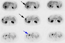
Comprehensive Cancer Network guidelines recommend FDG PET/CT in all patients with a disease stage of 1B2 or higher [26]. Because the cervix is in close proximity to the urinary bladder, FDG activity in the bladder may mask the primary, metastatic, and recurrent lesions [12]. For imaging patients with cervical carcinoma, a foley catheter and continuous bladder irrigation are useful to clear urinary activity [7,12,20]. Lasix also aids in clearing renal and ureteral activity [7]. Some authors also recommend a separate one-bed prone pelvic view to ensure separation of the bladder and pre-rectal space [20].
Primary lesion:
FDG is taken up by about 91% of primary cervical tumors [6]. Almost all primary cervical cancers greater than or equal to 7mm demonstrate FDG uptake [20]. Uptake has been correlated with over-expression of the Glut-1 glucose transporter [8]. Higher SUVmax values are associated with an increased risk of lymph node metastases [19]. High SUV max is also an independent predictor of recurrence after surgical treatment with or without adjuvant therapy [20]. The SUVmax value is also related to the likelihood for persistent disease following treatment and for overall survival [21]. Hence, cervical cancer patients with a high SUVmax may require more aggressive multimodality treatment [20].
Note that increased uptake can be seen within the endometrium above a cervical tumor and does not necessarily reflect tumor extension [9].
Nodal Metastases:
Lymphatic spread from cervical tumors is initially to the parametrial nodes [4,17]. From the parametrial nodes, the tumor may extend laterally to the external iliac nodes, to the hypogastric nodes that lie along the internal iliac vessels, or posteriorly along the uterosacral ligaments to the lateral sacral and sacral promontory nodes [4,17]. These latter three routes eventually drain into the common iliac nodes and subsequently para-aortic nodes [20]. Definition of lymph node status is one of the most important prognostic factors in cervical cancer [12]. For IB disease, survival is 85-95% for patients with negative nodes at surgery, but drops to 45-55% for those with positive nodes [2,4].
In one study comparing MR imaging with FDG PET, PET improved the
sensitivity in detection of lymph nodes metastases from 50-73% to
83-91% [6]. In a prospective evaluation of patients with clinical
stage IA or IB cervical cancer scheduled for treatment with
radical hysterectomy and pelvic lymph node dissection, PET/CT
showed a node based sensitivity of 72% for the detection of
metastatic disease (sensitivity was 100% for all nodes larger than
5 mm) [14]. Overall patient based detection of lymph node
involvement sensitivity is 73-77%, specificity 56-97%, PPV 92%,
NPV 89%, and accuracy 89% [14,17]. A
meta-analysis of PET/CT showed a pooled sensitivity of 79-84% and
a specificity of 95-99%; compared to 47-50% and 92-97%,
respectively for CT and 56-72% and 90-96%, respectively for MR
[20]. Overall PET/CT is more accurate than CT for evaluating lymph
node status and the overall sensitivity for the detection of lymph
node metastases increases with more advanced stage disease [21].
The risk of disease recurrence increases incrementally on the
basis of the most distant level of PET lymph node involvement
[22].
Para-aortic lymph node metastases:
Patients with paraaortic lymph node (PALN) metastases have a lower overall survival, disease free survival, and survival after recurrence [18,29]. Extended field radiotherapy is performed on patients whose disease has spread to PALN [29]. Despite this treatment, patients with PALN metastases have a substantially shorter median survival (33 months) and 40% of patients develop distant metastases [29]. The rate of paraaortic lymph node metastases is approximately 15-30% in patients with locally advanced cervical cancer [18]. Routine surgical staging of these nodes can have significant adverse effects and morbidities [18]. For the detection of paraaortic nodal metastases, both CT and MRI have low sensitivities (56% and 58%, respectively) [18].
In one study, metastatic para-aortic lymph nodes were identified with a sensitivity of about 82% [7]. However, in a meta-analysis, PET was found to perform acceptably only in patient populations at high risk for paraaortic nodal metastases- in patient populations with a prevalence of >15% PALN mets, the sensitivity was 73% [18]. But PET may be less accurate in patients with early stage disease as the overall sensitivity was only 34% [18]. In the same meta-analysis, the false positive rate was 27% (the false negative rate was 8%) [18]. It may be reasonable to omit surgical nodal exploration when both pelvic and paraaortic nodal uptake is present as these patients are much more likely to have metastatic disease [18].
Pelvic lymph node TLG and PALN SUVmax have been shown to be independently associated with overall survival [29]. It has been suggested that a PALN SUV max above 3.3 is significantly related to poor overall survival [29].
Findings on the PET exam can result in a change in planned treatment in 21% of patients [7]. Lymphoscintigraphy may aid in directing appropriate lymph node sampling when node dissection is to be performed in early grade patients [13].
Recurrent disease:
Higher FIGO stage is associated with an increased risk for recurrence [23]. Despite optimal initial treatment approximately 30% of cases of cervical cancer will recur following treatment for patients with stage IB or higher disease (10-20% for stages IB-IIA vs 50-70% for stages IIB-IV [23])- often with a combination of local and distant metastases [5,16,27]. The majority of recurrences occur within the first 2 years after completion of therapy- about 60% of recurrences occur within two years and 90% within 5 years [6,15]. Stage and lymph node status at the time of diagnosis are strong predictors of disease recurrence [15]. Patients with the greatest risk of recurrence are those with Stage IIb and Stage III cancer [5]. The most common sites for recurrence are the vaginal cuff, cervix, parametrium, and pelvic side wall [6]. Early detection of recurrence and institution of salvage treatment can result in improved patient survival [15]. However, curative therapy is futile in the presence of distant metastases [16].
PET/CT is highly accurate for the detection of locally recurrent or distant metastatic disease [21]. For the detection of recurrent cervical cancer, data indicate that FDG PET has a mean sensitivity and specificity of approximately 95% and 84%, respectively [10]. The mean diagnostic accuracy is about 88% [10]. Other authors suggest a pooled sensitivity of 87% and a specificity of 97% for distant metastases, and 82% and 98% for locoregional recurrence [26]. In a prospective evaluation of 249 patients with no evidence of cervical cancer following treatment, FDG PET imaging identified recurrence in 11% of patients [5]. The sensitivity and specificity of FDG PET for detection of early recurrence were 90% and 76%, respectively [5]. The sensitivity was lower for detection of lung, retrovesical nodes, and para-aortic nodes [5]. Overall, PET is superior to conventional imaging modalities for the detection of metastatic recurrent cervical cancer [11].
One critical issue in the evaluation of recurrent disease is distinguishing post-radiation changes from recurrent tumor which is not always possible with conventional imaging [6]. PET imaging can help to clarify the diagnosis of clinically suspected recurrence [16]. In patients with clinically equivocal recurrences FDG PET has a sensitivity of 92%, a specificity of 93%, a PPV of 96%, and a NPV of 87% [16]. PET findings can change the planed treatment in about 50% of cases [16]. In these patients, a positive PET also carried prognostic significance with overall decreased survival compared to patients with negative PET exams [16].
Distant metastases are seen in 70% of patients with recurrent disease - most frequently to the lung (21%), bone (20%- predominantly the pelvic bones, lumbar and thoracic spine), para-aortic nodes (11%), abdomninal cavity (8%), and supraclavicular nodes (7%) [20]. Patients with cervical adenocarcinoma are more likely to have evidence of adrenal and thoracic metastases than patients with squamous cell carcinoma [20]. The 10 year actuarial prevalence of distant metastases was ntoed to be 3% in stage IA, 16% in stage IB, 31% in stage IIA, 26% in stage IIB, 39% in stage III, and 75% in stage IV disease [20].
Another group of patients that can benefit from PET imaging are those with a prior complete response to therapy who present with rising tumor markers (squamous cell carcinoma antigen >1.5 ng/mL) and negative conventional imaging exams [13,20]. PET imaging can detect recurrent disease in up to 94% of these patients [13]. Early detection and early institution of appropriate therapy and lead to improved survival [13].
The findings on the PET exam can affect patient management in 23-65% of patients [10,16].
Prognosis/Monitoring response to therapy:
PET imaging is a strong predictor of disease-specific survival
[24]. FDG PET positivity for lymph node metastases correlates with
survival and is highly predictive of progression free survival
[20]. The risk of recurrent disease increases incrementally on the
basis of the most distant level of nodal involvement [24]. The 5
year survival rate has been reported to be 20% for patients with
common iliac or paraaortic metastases
on PET, compared to 85% for patients with isolated
pelvic nodal metastases [20]. Higher SUV max in metastatic lymph
nodes is also a significant negative prognostic factor in terms of
adverse events, persistence of disease after treatment, and
overall survival [23,29].
Higher SUVmax and higher volume based parameters, such as MTV and
TLG, have also been shown to be associated with a higher risk of
adverse events/recurrence (disease free survival), overall
survival, and death [23,25,26]. Higher levels of intratumoral
heterogeneity at initial staging are also associated with worse
overall survival [25].
Following primary treatment, PET imaging can provide additional
prognostic information compared to conventional imaging. A
complete metabolic response is associated with a very good
survival outcome [15]. FDG uptake at sites of primary treatment or
at new locations is a significant negative prognostic indicator
[10,15]. Two-year progression free
survival is 64% in CT-negative/PET-negative patients, 18% in
CT-negative/PET-positive patients, and 14% in
CT-positive/PET-positive patients [12]. In another study, 2 year
progression free survival was 80% in PET negative patients,
compared to only 40% in PET positive patients [12]. A prospective
study of patients with cervical cancer found a complete metabolic
response on PET imaging performed 8-16 weeks following completion
of therapy was associated with a 3 year progression free survival
of 78%; a partial metabloic response
with a survival rate of 33%; and the development of progressive
disease with a 0% survival rate [15]. Other authors report that
patients with no disease on a PET/CT exam performed 3-6 months
after chemoradiation have a 5 year survival rate of 92%, compared
to 46% for residual disease, and 0% if new areas of abnormal FDG
uptake are identified [22].
A 5-point qualitative scale has been developed to evaluate
treatment response [27]:
Score 1: No residual uptake (complete metabolic response)
Score 2: Focal uptake lower than mediastinal blood pool (likely CMR)
Score 3: Focal uptake above MBP, but less than liver (indeterminant)
Score 4: Focal uptake greater than liver (partial metabolic response)
Score 5: Focal uptake more than twice liver or new sites of disease (progressive disease)
Using this qualitative scoring system PET CT has been shown to
have a sensitivity of 94% and a specificity of 62% for predicting
residual or recurrent disease (PPV 59%, NPV 95%, and accuracy of
74%) [27]. A score of 1 or 2 has been shown to be associated with
95% disease free survival at 2 years, compared to a score of 5
which was associated with persistent or progressive disease in 93%
of patients [27]. There can be an overlap between partially
treated metabolic activity and mild-to-moderate inflammation
related FDG uptake (up to one-third of patients with a score of 3
are shown to have recurrent disease) [27]. Distinction between
focal versus diffuse uptake may help in differentiating
inflammation from recurrence [27].
Limitations of FDG PET imaging in cervical carcinoma:
Activity within the ureters may be misinterpreted as lymph node metastases [7]. False positive exams can be seen in nonmalignant processes such as endometriosis, infection, and benign tumors such as leiomyomas [21].
REFERENCES:
(1) Radiology 1998; Scheidler J, et al. Parametrial invasion in cervical carcinoma: Evaluation of detection at MR imaging with fat suppression. 206: 125-129
(2) Radiographics 2001; Panni HK, et al. CT evaluation of cervical cancer: spectrum of disease. 21: 1155-1168
(3) Radiographics 2003; Szklaruk F, et al. MR imaging of common and uncommon large pelvic masses. 23: 403-424
(4) AJR 2003; Kaur H, et al. Diagnosis, staging, and surveillance of cervical carcinoma. 180: 1621-1632
(5) J Nucl Med 2003; Ryu SY, et al. Detection of early recurrence with 18F-FDG PET in patients with cervical cancer. 44: 347-352
(6) AJR 2003; Kaur H, et al. Diagnosis, staging, and surveillance of cervical carcinoma. 180: 1621-1632
(7) J Nucl Med 2003; Ma SY, et al. Delayed 18F-FDG PET for detection of paraaortic lymph node metastatses in cervical cancer patients. 44: 1775-1783
(8) J Nucl Med 2004; Yen TC, et al. 18F-FDG uptake in squamous cell carcinoma of the cervix is correlated with glucose transporter 1 expression. 45: 22-29
(9) J Nucl Med 2004; Lerman H, et al. Normal and abnormal 18F-FDG endometrial and ovarian uptake in pre- and postmenopausal patients: assessment by PET/CT. 45: 266-271
(10) J Nucl Med 2004; Belhocine TZ. 18F-FDG PET imaging in posttherapy monitoring of cervical cancers: from diagnosis to prognosis. 45: 1602-1604
(11) J Nucl Med 2004; Yen TC, et al. Defining the priority of using 18F-FDG PET for recurrent cervical cancer. 45: 1632-1639
(12) Radiol Clin N Am 2004; Kumar R, Alavi A. PET imaging in gynecologic malignancies. 42: 1155-1167
(13) J Nucl Med 2005; Pandit-Taskar N. Oncologic imaging in gynecologic malignancies. 46: 1842-1850
(14) Radiology 2006; Sironi S, et al. Lymph node metastasis in patients with clinical early-stage cervical cancer: detection with integrated PET/CT. 238: 272-279
(15) JAMA 2007; Schwarz JK, et al. Association of posttherapy positron emission tomography with tumor response and survival in cervical carcinoma. 298: 2289-2295
(16) J Nucl Med 2008; van der Veldt AAM, et al. Clarifying the diagnosis of clinically suspected recurrence of cervical cancer: impact of 18F-FDG PET. 49: 1936-1943
(17) AJR 2009; Pandharipande PV, et al. MRI and PET/CT for triaging stage IB clinically operable cervical cancer to appropriate therapy: decision analysis to assess patient outcomes. 802-814
(18) J Nucl Med 2010; Kang S, et al. Diagnostic value of 18F-FDG PET for evaluation of paraaortic nodal metastasis in patients with cervical carcinoma: a metaanalysis. 51: 360-367
(19) AJR 2010; Kitajima K, et al. Spectrum of FDG PET/CT findings of uterine tumors. 195: 737-743
(20) Radiographics 2010; Son H, et al. PET/CT evaluation of cervical cancer: spectrum of disease. 30: 1251-1268
(21) AJR 2011; Patel CN, et al. 18F-FDG PET/CT of
cervical carcinoma. 196: 1225-1233
(22) J Nucl Med 2015; Lee SI, et al. Evaluation of gynecologic
cancer with MR imaging, 18F-FDG PET/CT, and PET/MR
imaging. 56: 436-443
(23) AJR 2018; Han S, et al. Prognostic value of volume-based
metabolic parameters of 18F-FDG PET/CT in uterine
cervical cancer: a systematic review and meta-analysis. 211:
1112-1121
(24) Radiology 2019; Lee SI, Atri M. 2018 FIGO staging system for
uterine cervical cancer: enter cross-sectional imaging. 292: 15-24
(25) AJR 2020; Pinho DF, et al. Value of intratumoral metabolic
heterogeneity and quantitative 18F-FDG PET/CT
parameters in predicting prognosis for patients with cervical
cancer. 214: 908-916
(26) AJR 2020; Mansoori B, et al. Multimodality imaging of
uterine cervical malignancies. 215: 292-304
(27) AJR 2020; Banks KP, et al. It's about quality, not quantity:
qualitative FDG PET/CT criteria for therapy response assessment in
clinical practice. 215: 313-324
(28) Radiographics 2020; Salib MY, et al. 2018 FIGO staging
classification for cervical cancer: added benefits of imaging. 40:
1807-1822
(29) J Nucl Med 2020; Leray H, et al. 18F-FDGPET/CT identifies predictors of survival in patients with locally advanced cervical carcinoma and paraaortic lymph node involvement to allow intensification of treatment. 61: 1442-1447
