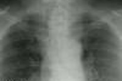
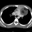
Invasive Thymoma:
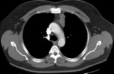
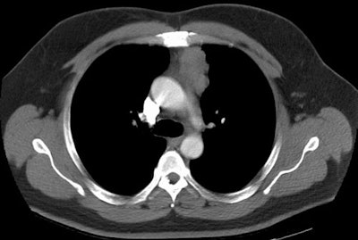
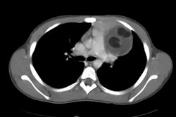
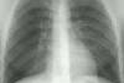
The images below demonstrate an invasive thymoma. The first image demonstrates an eccentric, lobulated anterior mediastinal mas. There is ill-defined increased density within the anterior mediastinal fat, as well as the presence of a borderline enlarged mediastinal lymph node adjacent to the mass. On the lower image, the mass lacks a well defined border and again noted is infiltration of the mediastinal fat. These features suggest an invasive thymoma. (Click on images to enlarge)
