If you've been in radiology long enough, chances are you've seen your share of strange cases. But radiologists at a Southern California hospital got a doozy in April, when a construction worker presented with obvious head injuries but no visible sign of their cause.
Dear AuntMinnie Member,
If you've been in radiology long enough, chances are you've seen your share of strange cases. But radiologists at a Southern California hospital got a doozy in April, when a construction worker presented with obvious head injuries but no visible sign of their cause.
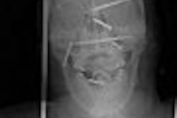
Imaging studies subsequently indicated the diagnosis: Six nails had been driven into the man's head by a nail gun that had fired accidentally. The radiologists made the initial diagnosis with radiography, then used 3D CT to fine-tune their findings and help prepare the man for surgery, according to an article we're featuring this week in our CT Digital Community by staff writer Tracie L. Thompson.
Radiologists reading the 3D reconstructions were able to demonstrate the location of the nails in relation to crucial brain anatomy, such as major arteries. The CT images, used in conjunction with a subsequent angiogram, helped surgeons remove the nails, and the man eventually recovered from the incident.
See the images for yourself in the CT Digital Community, at ct.auntminnie.com.


