Iso-osmolar contrast agents do not offer significant advantages over low-osmolarity agents in arterial enhancement quality in CT angiography studies, a study published online in Radiology has concluded.
"We believe enhancement quality alone should not be considered an indication for the preference of iso-osmolar over low-osmolarity agents in that specific clinical setting," wrote researchers from the department of radiology at the Centre Hospitalier Universitaire de Sherbrooke in Quebec, Canada.
The conclusions came at the end of a five-month study involving 47 patients, in which 188 pulmonary arterial branches were examined and evaluated for contrast enhancement.
The findings are significant because of the increasing role of pulmonary CT angiography in diagnosing pulmonary embolism, and the role of contrast agents in the enhancement quality of the pulmonary arteries.
Iso-osmolar versus low-osmolarity agents
The newer nonionic iso-osmolar agents are more expensive than low-osmolar agents, but the higher price could be offset by an improved safety profile. Recent research has indicated that iso-osmolar contrast is less likely to be nephrotoxic in high-risk patients compared with low-osmolarity agents (New England Journal of Medicine, February 2003, Vol. 348:6, pp. 491-499).
Studies have also shown the rate of injection-related pain is also lower with iso-osmolar agents (CardioVascular and Interventional Radiology, July 1997, Vol. 20:4, pp. 251-256). The viscosity of iso-osmolar agents is the same as that of blood, and therefore, theoretically less prone to dilution than the thicker low-osmolarity agents.
But these advantages apparently don't translate into better images. The researchers from Canada found there were no significant benefits in enhancement quality between the two contrast agents for pulmonary CT angiography.
The reason may be that arterial enhancement studies require a shorter lag time between injection and imaging, and the dilution effect related to osmolarity of contrast agents is not as apparent, according to the study authors.
"These results agree with those in studies in which iso-osmolar and low-osmolarity contrast agents were compared for arterial enhancement," they wrote (Radiology, January 28, 2005).
The iso-osmolar contrast agent used in the study was iodixanol (Visipaque, GE Healthcare Bio-Sciences, Little Chalfont, U.K.), and the low-osmolarity agent was iohexol (Omnipaque, GE Healthcare Bio-Sciences). Of the 47 patients in study, 25 were administered iodixanol and 22 received iohexol.
"The volume of contrast injected was calculated to provide an equal quantity of iodine. One hundred milliliters of a low-osmolarity contrast agent, iohexol, or 90 mL of an iso-osmolar contrast agent, iodixanol, was injected, with a total of 24 or 24.3 g of iodine, respectively," the researchers stated.
CT angiography
CT angiography was performed with a four-slice Aquilion CT scanner (Toshiba Canada, Kirkland, Canada) according to a standard single-breath-hold protocol (200 mA, 100 mAs, 120 kV).
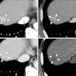
Imaging was done from the pulmonary apex to the diaphragm, and scans were acquired in a craniocaudal direction. The quality of vascular enhancement was evaluated at four designated levels of the pulmonary arteries of the lower lobe of the left lung, the lobar artery, one segmental (posterobasal) artery, and two subdivisions of this segmental artery (posterior and mediobasal ramus).
The lower lobe was chosen because a majority of pulmonary emboli are found here. The left lung was chosen because it has more anatomic variation in the pulmonary tree compared to the right lung. In cases in which an embolus was identified in the left lung, the equivalent arteries in the right lung were evaluated.
Evaluation of vascular enhancement
Evaluation of vascular enhancement was done by three radiologists blinded to each other's interpretation. The enhancement was assessed as "good to excellent" if it allowed confident diagnosis of the presence or absence of a clot. It was assessed as "limited but of diagnostic value" if the enhancement was not optimal but sufficient to allow diagnosis. The ranking "not diagnostic" was used if the enhancement was inadequate for diagnosis.
Homogeneity of vascular enhancement was also graded on a scale from 1 to 3 using a template obtained from CT angiographic examinations performed prior to the study.
"Quality of enhancement of lobar and segmental arteries was less frequently considered 'not diagnostic' in the patients who received iodixanol than it was in the patients who received iohexol, but the difference was not statistically significant (p > 0.05)," the researchers reported.
"On the other hand, quality of enhancement of subsegmental arteries of the second order more often was considered 'good to excellent' in patients who received iohexol than it was in patients who received iodixanol when we used data from reader 1 (p < 0.05) and mode data (p < 0.1)," they added.
There were no other significant differences between the two groups, according to the study.
By N. Shivapriya
AuntMinnie.com contributing writer
February 3, 2005
Related Reading
Iso-osmolar contrast agent less likely to be nephrotoxic in high-risk patients, February 6, 2003
Copyright © 2005 AuntMinnie.com


