BERLIN - A principal limitation of lung cancer imaging is the difficulty of distinguishing lung nodules attached to blood vessels from normal blood vessels.
Such nodules are especially hard to nail down in CT data when they are malignant, in which case they are even likelier to infiltrate pulmonary blood vessels surreptitiously -- rather than displace them in visually obvious ways, as benign granulomas often do.
On the other hand, malignant nodules attached to blood vessels, unseen and well supplied with blood, can be fast-growing and deadly.
But the stealth nodules have a new enemy in Dr. Sumiaki Matsumoto, Ph.D., and colleagues from the Kobe Graduate School of Medicine and Toshiba Medical Systems in Japan. The group developed a 3D feature they call the diminution index, which scans the CT pulmonary blood vessel data for signs of rapidly expanding shapes that can indicate a nodule rather than a blood vessel. If blood vessel diameter "diminishes" too abruptly, the researchers say, it is usually a nodule.
A CAD system based on the diminution index, along with five auxiliary features, found most of the vessel-contiguous simulated nodules it was looking for, yielding a moderate false-positive rate and reasonably good sensitivity, Matsumoto said in a discussion of his group's data. Pulmonary blood vessels are the largest source of false positives in CT lung exams, he said at the thoracic CAD session of the 2005 Computer Assisted Radiology and Surgery (CARS) meeting.
"A key problem in computer-aided lung nodule detection of CT images is how to suppress false-positive images due to blood vessels without compromising sensitivity," Matsumoto explained. "Because lung nodules can be attached to pulmonary blood vessels, this problem boils down to how to differentiate between pulmonary blood vessels and nodules attached to blood vessels."
The diminution index aims to attack this problem. It is derived from a 3D region obtained by exploring the candidate nodule and its surroundings in a centrifugal direction with respect to the nodule, measuring the diminution of this 3D region in the centrifugal direction. If the nodule candidate is part of a blood vessel, its diminution index can be expected to be small or nonexistent. If it is a nodule, its diminution index should be large, according to the hypothesis.
The team acquired 16 "almost normal" CT datasets obtained on a 16-detector Aquilion scanner (Toshiba Medical Systems, Tokyo), and added 130 simulated nodules measuring 3-5 mm in diameter near the blood vessels. The acquisition parameters included 120 kVp, 300 mAs, and 16 x 0.5-mm collimation. The simulated nodule data were created from small regions of interest on blood vessels.
Matsumoto explained the complex process that leads to calculation of the diminution index. First nodule candidates are extracted using deformable ellipsoid models by segmenting the lung region in the CT data via adaptive thresholding, he said. A distance transform method is then used to create an ellipsoid model of the candidate nodule, whose boundaries and connected components are determined by means of gray levels. The diminution index is calculated from the center of the ellipsoid in this region using a region-growing method.
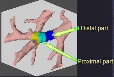
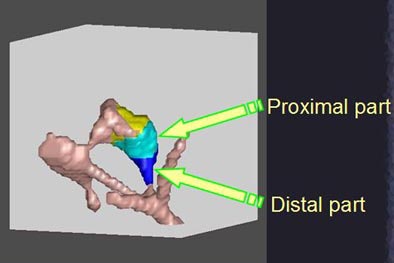
"The region growing is carried out over a distance proportional to the size of the bound ellipsoid," Matsumoto said. "The distance is set to twice the radius of the bounding ellipsoid in the centrifugal direction. Then the 3D region is divided into its proximal and distal parts by the bounding ellipsoid."
The diminution index is calculated as 1 minus the ratio of the volume of the distal part divided by the volume of the proximal part, Matsumoto said.
"There are multiple centrifugal directions to be considered, so the formula is computed for each direction," he said. "The diminution index is defined as the minimum value of the numeric."
The rest of the CAD scheme qualifiers are standard, including, for example, the diameter of the nodule and contrast with respect to the surrounding tissues, Matsumoto said. A subset of data with 30 nodules was used to train the system, and the remaining 100 nodules were actually used to test the CAD scheme.
 |
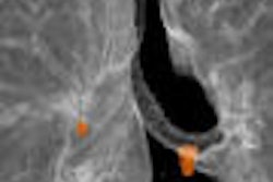
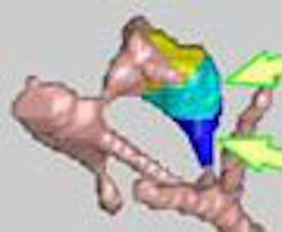
| A small diminution index of 0.11 in the image above is associated with normal blood vessels. A larger diminution index of 0.78 in the image below is associated with a nodule attached to a blood vessel. All images courtesy of Dr. Sumiaki Matsumoto, Ph.D., and Hitoshi Yamagata, Ph.D. |
 |
The results curve below was obtained by varying the cutoff values of the definition index, he said. The results showed 100% sensitivity for detection of the nodules with a high false-positive rate of 60. As the cutoff value of the index increased, the number of false-positives was reduced. A representative point was 78% sensitivity with 5.3 false-positives per case.
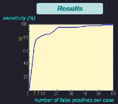 |
| The results showed good sensitivity with a reasonable number of false positives. As the cutoff value of the diminution index increased, the number of false positives was reduced. A representative point can be seen at 78% sensitivity with 5.3 false positives per case. |
The index provides a natural means of differentiating pulmonary blood vessels from nodules attached to vessels, Matsumoto said.
Unfortunately, the thicker hilar vessels frequently caused false positives, he noted, showing an example of a vessel structure with an inappropriately high diminution index. The index, which relies on the gradual tapering of blood vessels, can be misleading in the hilar vessels because they decrease rapidly in size as they branch out of the mediastinum. This problem is a focus of the group's current research, he said.
"We have introduced a novel 3D feature, the 'diminution index,'" Matsumoto said. "Although imperfect, it has the capability to differentiate between pulmonary (blood vessels) and nodules attached to vessels, and seems promising for the detection of lung nodules in CT images."
By Eric Barnes
AuntMinnie.com staff writer
June 26, 2005
Related Reading
CAD substantially improves MDCT's sensitivity in detecting lung nodules, January 12, 2005
Lung CAD makes strides in nodule detection, January 30, 2004
Lung CAD applications starting to outread radiologists, February 13, 2003
Copyright © 2005 AuntMinnie.com


