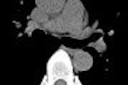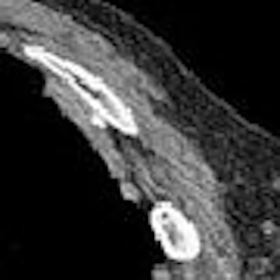
As the debate swirls in the U.S. over CT lung screening of smokers, Canadian researchers are developing a unique CT screening program designed to focus on patients who have been exposed to asbestos as a tool for early detection of malignant mesothelioma.
Researchers from University Health Network and Princess Margaret Hospital in Toronto are looking for cases of lung cancer and malignant mesothelioma in their cohort of patients who have had long-term exposure to asbestos fibers. The investigators aim to help patients while advancing the understanding of mesothelioma, especially early-stage disease, which is poorly understood.
"We have developed a program for screening with low-dose CT using prior exposure to asbestos as a risk factor," said Dr. Heidi Roberts in a presentation at the 2008 European Congress of Radiology (ECR) in Vienna. "Asbestos exposure is a risk factor for the development of mesothelioma, and, in addition, the disease has a marker: pleural plaques are known as a reliable marker for prior asbestos exposure."
Like most disease processes, prognosis is favorable in patients diagnosed early. However, current treatments are radical, and questions remain about the appearance of early mesothelioma, Roberts said.
Roberts and colleagues, including Drs. Michael Johnston and Brenda O'Sullivan, began the study three years ago, enrolling 424 subjects (416 men, eight women; ages 32-83 years, mean age 61) at risk for mesothelioma.
"Risk was defined as known and documented prior asbestos exposure at least 20 years ago. If they did not know about prior asbestos exposure, (risk) would be the presence of pleural plaques documented on chest radiographs," Roberts said. Most of the cohort (78%) had a history of smoking; 22% had never smoked.
All subjects were screened with low-dose protocols on multidetector-row CT (MDCT) scanners equipped with four to 64 detector rows. Patients were scanned at 120 kVp and 40-60 mAs, and 1.25-mm overlapping reconstructions were used to examine the data.
Most patients with inconspicuous pleural plaques and/or small, indeterminate lung nodules were scheduled for one-year follow-up. Those with suspicious lung nodules were referred for a three-month follow-up or immediate biopsy if warranted.
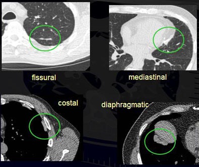
The results showed that 308 (73%) patients had pleural plaques. At least 83% of the plaques had at least some calcification, ranging from "tiny spots" at CT to complete calcification in 11%. Most involved the costal (more than 97% of plaques) and diaphragmatic (more than 88% of plaques) pleura.
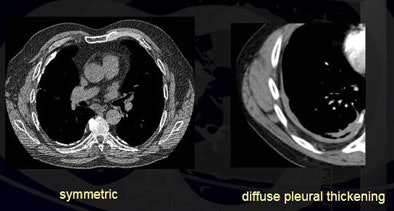
In all, 86% of the plaques were flat and 91% were symmetrical. More than 80% of pleural plaques "were completely flattened and inconspicuous, while 10% had these lobulated and rather thick nodules," Roberts noted.
Conversely, 14% of plaques were lobulated, 2% were asymmetric with a right-sided dominance, and 1% were associated with pleural effusions. Eleven percent of patients had diffuse pleural thickening.
In all, 312 (75%) had pulmonary nodules, and a total of 103 nodules in 86 subjects were larger than 5 mm in diameter, Roberts said. Two other biopsies yielded insufficient material, and the nodules are currently regarded as benign while the subjects remain under surveillance.
 |
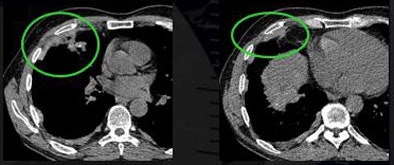 |
| Researchers found that more than 86% of pleural plaques were completely flat and 91% were symmetrical. Eleven percent of patients had diffuse pleural thickening (top). Plaques that were asymmetrical in shape and mass-like were considered suspicious for malignancy (above). Plaques were detected in the lung fissures, mediastinum, diaphragm, and costal regions (below). All images courtesy of Dr. Heidi Roberts. |
 |
In addition, the researchers found two malignant pleural mesotheliomas and one malignant peritoneal mesothelioma, for an overall malignancy rate of 1.4% to 1.9%.
Malignancies
The incidence of cancer was actually lower than in the institution's similar study of smokers "because we included nonsmokers -- and we found more lung cancers than pure mesotheliomas, which we expected," Roberts explained.
The single-center study "did detect lung cancers, both early- and late-stage ones, and we also found mesothelioma, so far only late-stage," she said, noting that many patients are scheduled within the next couple of months for their one-year follow-up, which is expected to reveal new information about disease progression.
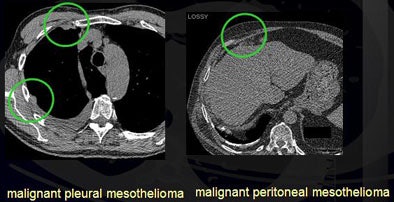 |
| Detected malignancies included malignant pleural mesothelioma and malignant peritoneal mesothelioma. |
Some treatments are available for malignant mesothelioma, most involving quite radical therapies such as chemotherapy, pneumonectomy, and radiation, "but they do have quite promising results," Roberts said.
An audience member asked how early-stage mesothelioma is defined and how it appears on CT images.
"That's the whole problem. Nobody really knows what early-stage mesothelioma looks like," she said. "The literature talks about mesothelioma a lot, but it's mainly about staging and disease management. So hopefully, in the next year, when we have a lot of annual follow-ups analyzed and scanned, we'll be able to answer this question."
With the team's well-funded screening program comes hope that knowledge of treatment and disease management will advance fast enough for patients to benefit from the results, Roberts added.
"This is exactly where we were for a couple of decades ago with lung cancer, where we were screening for lung cancer, but we couldn't do anything about it anyway," Roberts said. "And look where we are now. We have (such) early lung cancer detection that even staging regimens have to be redefined. Hopefully, there will be a parallel development where we can find the early mesothelioma, and the early stage of disease can be treated."
By Eric Barnes
AuntMinnie.com staff writer
April 3, 2008
Related Reading
Workplace cancers cause 200,000 deaths a year: WHO, April 30, 2007
Men show higher malignant mesothelioma rates after asbestos exposure, March 2, 2007
CT distinguishes asbestos lung damage from smoking impairment, January 10, 2007
Indian shipbreakers suffering from asbestos -- report, September 9, 2006
Report links asbestos to larynx cancer, June 8, 2006
Copyright © 2008 AuntMinnie.com


