Newly introduced scanners don't come with optimized imaging protocols for every application. It's up to researchers, like cooks in a kitchen without a recipe book, to figure out what works and what doesn't.
Take the new 320-slice CT scanner (AquilionOne, Toshiba America Medical Systems, Tustin, CA), unveiled at last year's RSNA meeting. Until now, heart imaging with the wide-area-detector machine has focused mainly on retrospectively gated CT angiography (CTA).
Prospective gating, a technique that has gained rapid traction for its ability to trim dose in 64-slice CTA applications, can theoretically do the same for 320-slice imaging. But researchers had yet to optimize settings and minimize patient dose for 320-slice prospectively gated CTA.
In a new study, Dr. Michael Steigner, along with Dr. Hansel Otero, Dr. Frank Rybicki, and colleagues from Brigham and Women's Hospital in Boston, did so by evaluating the impact of different amounts of "padding," or phase window width, on image quality and dose in prospectively gated CTA (International Journal of Cardiovascular Imaging online, July 29, 2008).
"The purpose of this study is to study the relationship between the phase window width and image quality for single RR acquisition coronary CTA, and to study the relationship between heart rate and the width of the phase window with diagnostic quality images," Steigner and colleagues wrote.
 |
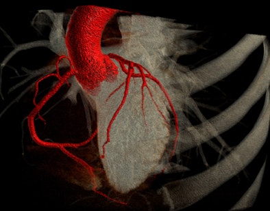
| 3D volume rendering performed on 320-detector-row MDCT scanner. All images courtesy of Dr. Michael Steigner. |
Electrocardiogram (ECG) gating synchronizes the CT data acquisition with the cardiac cycle, and the resulting images are reconstructed during the cardiac phases with minimum motion, typically mid- to end-diastole. With retrospective gating, images are acquired throughout the cardiac cycle, and the reconstruction phase with the least motion artifact is chosen.
 |
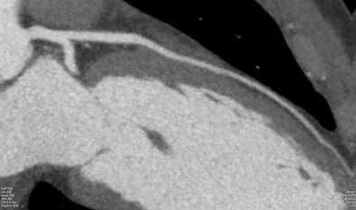
| Curved multiplanar reformat of the left anterior descending (LAD) artery performed on the 320-detector-row MDCT scanner. |
The phase window is the period of the cardiac cycle during which the tube current is activated, the group explained. It is closely associated with dose, because a wider window corresponds to a greater exposure. Therefore, the optimal phase window should be wide enough to include a range of optimal reconstruction phases without having to rescan the patient, but small enough to minimize dose, the researchers explained.
Another consideration: The phase with the least motion artifact is often different for the left and right coronary artery systems, especially in subjects with higher heart rates.
The AquilionOne's wide 16-cm coverage enables the heart to be scanned in a single beat, eliminating the need to reconstruct images from several heartbeats and the resulting image artifacts, while reducing helical oversampling and, one hopes, the dose to the patient.
But new tools were needed to evaluate radiation dose on the new system. "Wide-area-detector CT dosimetry is still an active topic of investigation," they noted. "Standard [CT dose index (CTDI)] methods currently in practice for 64-slice CT scanners do not directly apply since the wide-area-detector primary x-ray beam is larger than both the standard ionization chamber and the dosimetry phantom," Steigner and his colleagues wrote.
The Brigham study
The Brigham researchers tested their approach in a population of 41 consecutive patients (mean age 53, range 47-79), of whom 34 were being evaluated for chest pain and seven for presurgical planning. All patients were scanned using 320-slice CTA with prospective gating and a wide 60%-95% phase window. A scaling factor based on a 300-mm chamber combined with a 360-mm-long dosimetry phantom was used to estimate effective dose.
Two experienced readers then effectively "narrowed" the window by retrospectively looking for the best reconstruction phase within the data for the left anterior descending (LAD) artery, left circumflex (LCx) artery, and right coronary artery (RCA) within each dataset.
Before scanning, all patients received 80 mL iopamidol iodinated contrast agent (Isovue-370, Bracco Diagnostics, Princeton, NJ) and 40 mL saline, both injected at 6 mL/sec (Empower CTA dual injector, E-Z-EM, Lake Success, NY). Beta blockers were administered intravenously (metoprolol, 5 mg increments) to achieve target heart rates of 65 bpm. Finally, all patients received 0.4 mg sublingual nitroglycerine.
The subjects were scanned at 320 x 0.5-mm detector configuration, at 120 kVp and 400 mA, while 14 larger patients (> 30 kg/m2) were scanned at 580 mA. The gantry rotation time was 350 ms.
"For larger patients, lowering the dose via mAs reductions is often ineffective since the resultant images have a decreased signal-to-noise ratio," the authors wrote. "The radiation dose is linearly related to the mAs and craniocaudal imaging field-of-view. The relationship between effective dose, kV, and the prospective phase window width is more complex."
The first method to assess the influence of varying the phase window width was to consider individual coronary arteries plus the combination of the LAD, LCx, and RCA with at least one diagnostic phase for the following window widths: 75% alone, 70%-79%, 60%-79%, and 60%-95% (the entire acquisition). The second analysis estimated the smallest phase window width that includes at least one diagnostic phase for 95% of coronary arteries, they explained.
The 41 patients included 22 with normal coronary arteries, 12 with coronary artery disease but no stenosis greater than 50% in diameter, and seven with one or more stenotic lesions greater than 50% in diameter.
Widening the phase windows increased the proportion of coronary arteries with at least one diagnostic phase, the group reported. Among the 41 patients, 95% of vessels had a diagnostic phase in the 72%-77% phase window. After accounting for sampling variation, the 72%-81% phase window was shown to have a 0.95 probability of including a diagnostic phase for 95% of coronary arteries in the general population.
The 72%-81% phase window included 98% of the vessels in the sample. Interobserver agreement between the two readers was 0.959, with a 0.95 confidence interval (0.908, 0.987). Patients with lower heart rates had significantly more diagnostic phases.
"This patient cohort had an estimated mean effective dose of 6.7 mSv," Steigner and colleagues reported. "For these patients, had the phase window width been 1%, 10%, or 20%, the estimated mean effective dose would have been 4.1, 5.3, and 6.5 mSv, respectively."
For prospectively ECG-gated single-heartbeat coronary CTA, a phase window width of 10% will reduce patient radiation and yield diagnostic images in more than 90% of patients, they wrote. Heart rate control is key to minimizing dose because it increases confidence that a narrower and thus lower-dose phase window will yield diagnostic images, they added.
"The limit of narrowing the phase window width to only 75% is expected to yield diagnostic images for the entire coronary tree in approximately 65% of patients," they wrote. "Increasing the window width to 10% will include greater than 90% of patients, with little change between 10% and 20%. Thus, we recommend a standard window width of 10% for single-heartbeat 320-detector-row coronary CTA, noting that the decision should be made on a per-patient basis since patients with a low heart rate can likely be imaged at lower dose."
Larger studies are needed to fine-tune the clinical guidelines for choosing the best phase window width for 320-detector-row coronary CTA.
"The conservative initial approach to prospectively gated single RR 320-slice coronary CTA lends itself to retrospective analyses of dose reduction via narrowing the phase window," they wrote. "For prospectively ECG-gated single-heartbeat coronary CTA, a phase window width of 10% will reduce patient radiation and yield diagnostic images in greater than 90% of patients. Heart-rate control is an important component of 320-detector-row prospectively gated CT dose reduction."
By Eric Barnes
AuntMinnie.com staff writer
August 5, 2008
Related Reading
AHA urges cautious use of coronary CTA and MRA, July 9, 2008
Fourth-generation MDCT scanners do cardiac differently, July 8, 2008
First 320-slice coronary CTA study shows high image quality, April 7, 2008
AquilionOne 320-slice CT scanner spearheads Toshiba RSNA launches, November 26, 2007
Radiation dose slashed in 64-slice coronary CTA, February 15, 2007
Copyright © 2008 AuntMinnie.com


