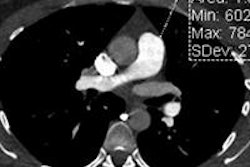
A series of studies and an expert panel discussion published in the American Journal of Roentgenology highlight the benefits and risks of CT exams for detecting suspected pulmonary embolism (PE). On the whole, they downplayed the notion that the modality is overused.
CT angiography (CTA) scans for PE typically achieve low positivity rates even in the most carefully selected patient populations, but risks can be minimized by keeping radiation doses low and by doing a careful risk-benefit analysis before the scan, the authors reported.
What was their take-home message? Pulmonary CTA for PE isn't perfect, but its many benefits include a look at the heart -- specifically, the right ventricle -- for extrapulmonary problems. Among the drawbacks, CT is identifying more nonfatal cases of PE than ventilation/perfusion (V/Q) scans did, leading to questions of overdiagnosis. And in the final analysis, there is no plausible replacement for CT on the horizon, according to the authors.
This article provides summaries of three of the five studies included in the June issue (Vol. 198:6) of AJR, as well as a few comments from a panel discussion featuring the authors.
CT scan benefits outweigh risks
Depending on multiple factors such as patient demographics and radiation dose, the benefits of using CT for suspected PE range from 25 to 187 times the risks, wrote Dr. James Woo and colleagues from Vancouver General Hospital (AJR, pp. 1332-1339).
Woo's group looked at the risks and benefits of pulmonary embolism screening in 1,424 patients, noting the exam setting, patient age, sex, and interpretation results for PE, as well as the radiation exposure, estimating the lifetime risk of cancer using the 2006 Biological Effects of Ionizing Radiation (BEIR) VII report.
Benefit-to-risk ratios were calculated by dividing the mortality benefit of preventing fatal PE by the risk of radiation-induced cancer from the scan.
PE was found in 188 (13.2%) of the 1,424 patients. Men received significantly higher radiation doses (9.7 mSv) than women (8.4 mSv), but they still had a lower lifetime attributable risk of cancer mortality (p < 0.0001) than women.
PE burden no predictor of mortality
In another study, Dr. Michael Morris and colleagues from the Mayo Clinic in Scottsdale, AZ, and Rochester, MN, examined CT findings and long-term mortality after PE, which is unknown (AJR, pp. 1346-1352).
The study examined whether three CT findings -- increased embolic burden, interventricular septal bowing toward the left ventricle, and right-to-left ventricle diameter ratio greater than 1 -- are independent predictors of long-term all-cause mortality after PE.
In scans reviewed by three observers, CT findings and clinical information were compared for all-cause mortality. The median survival was 6.2 years after acute PE diagnosis, with an estimated 10-year survival rate of 37.4%.
CT-derived embolic burden was associated with only a very small decrease in long-term all-cause mortality in both univariate (hazard ratio [HR], 0.97; p < 0.001) and multivariate (HR, 0.97; p < 0.001) analyses, the group reported. In addition, interventricular septal bowing and right-to-left ventricle diameter ratio were not significantly associated with long-term all-cause mortality.
The results may seem paradoxical, the group wrote, but similar findings have been noted in other studies.
"Patients with massive PE who survive to undergo CT may represent a unique subset of patients who have an improved overall survival, as opposed to those with massive PE who die before imaging," they wrote. "Alternatively, the relationship between embolic burden and survival may be due to differences in treatment not accounted for in our study."
Future studies may be able to clarify the findings by examining CT exams beyond the initial scan at presentation.
Does CT overdiagnose?
CT appears to diagnose more mild, nonfatal cases of PE than V/Q scintigraphy, according to another study (AJR, pp. 1340-1345).
Dr. Steven Sheh and colleagues from Albert Einstein College of Medicine investigated whether pulmonary emboli diagnosed with pulmonary CTA "represent a milder disease spectrum than those diagnosed with ... V/Q scintigraphy," in a study that also looked at incidence and mortality trends among patients diagnosed with PE over a seven-year period.
Over the seven years, 2,087 patients (1,361 women, 726 men; mean age, 61.8 years) with PE were identified. Sheh and colleagues used logistic regression analysis to estimate the odds of death for PE diagnosed with pulmonary CTA versus PE diagnosed with V/Q scintigraphy.
From 2000 to 2007, the incidence of pulmonary embolism increased from 0.69 to 0.91 per 100 admissions; this strongly correlated to increased use of CT over the seven-year period. Mortality was unchanged, but the case-fatality rate decreased from 5.7% to 3.3% over the same time period, the group reported.
Cases of PE diagnosed with pulmonary CTA were, on average, half as lethal as those diagnosed with V/Q scintigraphy (odds ratio, 0.538; 95% confidence interval: 0.314-0.921), Sheh and colleagues wrote.
The results are evidence that a shift in imaging from V/Q scintigraphy to pulmonary CTA increased the diagnosis of a less fatal spectrum of PE, "raising the possibility of overdiagnosis," they stated.
"Because current treatment recommendations were designed before the widespread adoption of pulmonary CTA, outcome-based clinical trials with long-term follow-up would be helpful to further guide management," the authors wrote.
Panel discussion
The AJR issue also included a panel discussion that fielded questions regarding the findings of the five studies (pp. 1313-1319).
Addressing a question on the risks versus benefits of CTA, Dr. Philip Araoz from the Mayo Clinic in Rochester, MN, noted that the risks of PE far outweigh those of CT scans.
"The mortality rate associated with untreated PE is not well-known but is often reported to be 30%," he wrote. "By comparison, risks of radiation at the level of diagnostic CT have never been detected. All reports of this radiation risk are hypothetical, but even assuming that these hypothetical risks are accurate, they amount to a less than 1% increased incidence of cancer mortality for the highest risk group (i.e., young women)."
Patients undergoing pulmonary CTA tend to be older and at even lower risk of carcinogenesis due to radiation, Araoz wrote. For example, in the Morris et al study, the mean age of the patients undergoing pulmonary CTA was 63 years.
The risks of diagnosing and treating PE must be compared to the risks of nontreatment, added Dr. Eduardo Barbosa Jr., from the Hospital of the University of Pennsylvania. The risks from not diagnosing or treating PE are difficult to estimate as they depend on several factors, including the "pretest probability of PE, the likelihood of alternative treatable conditions, the patient's comorbidities, and the clot burden."
The current risks of nontreatment have dropped from about 30% in the 1940s to perhaps 5% today, but they vary by case and are subject to debate, according to Barbosa.
Overdiagnosing and overtreating PE?
The panel also addressed the question of overdiagnosis, i.e., whether physicians are diagnosing and treating cases with a low pretest probability of PE (probably), and whether some cases should be followed rather than treated (probably not).
"It is true that the number of pulmonary CTAs is increasing rapidly and the percentage of positive scans is decreasing," Araoz wrote. "However, the rate of positive pulmonary CTAs is still in the range of 5% to 10% in most studies, which may be reasonable considering that PE is difficult to diagnose clinically and is potentially life threatening when untreated."
As for whether some PEs can be followed, the answer is difficult to determine because current practice is to treat all PEs with anticoagulation, he wrote.
Addressing the question of whether MRI or another modality could potentially replace CT, Araoz noted that MRI allows for pulmonary perfusion imaging, and when used in combination, it becomes a powerful diagnostic tool.
"However, a large percentage of patients undergoing pulmonary CTA have significant lung findings, which will always make MRI less attractive than CT for diagnosis of PE," he wrote.
Barbosa added that MRI and MR angiography can detect central PE with accuracy equivalent to that of pulmonary CTA; however, pulmonary CTA is superior for segmental and subsegmental arteries, he wrote.
Finally, Barbosa said that V/Q cannot be competitive with CTA for PE assessment. V/Q is not inferior to CTA for diagnosis, he said, but it is less sensitive and not as accurate in quantifying the embolic burden.
V/Q "is limited in the setting of extensive pulmonary parenchymal abnormalities that cause matched V/Q defects and it cannot assess the pulmonary parenchyma or the [right ventricle], which are important predictors of outcomes," he wrote.


