A new coronary artery segmentation and tracking method is showing promise for supporting computer-aided detection (CAD) schemes that can read coronary CT angiography (CCTA) scans to look for arteries blocked by both soft and calcified plaque, according to research published in Medical Physics.
The technique, developed as a critical first step in a CAD scheme, is highly sensitive for segmenting and tracking the coronary arteries, according to the authors from the University of Michigan. Marks generated by the algorithm overlapped with more than 80% of coronary artery center points, and the scheme detected more than 90% of marks left by radiologists who were working without the aid of any software, the group reported.
The algorithm's false-positive rate turned out to be less than one per case, lead author Chuan Zhou, PhD, told AuntMinnie.com in an interview. Zhou is an associate research professor in the university's radiology department (Med Phys, August 2014, Vol. 41:8).
More slices to review
Rapid technical advances in CT have made CCTA one of the most important modalities for evaluating coronary atherosclerosis. But more detector rows mean more CT image slices to review, and with patient volumes growing, radiologists evaluating CCTA are finding that they need help to examine the blood vessels adequately.
 Chuan Zhou, PhD, from the University of Michigan.
Patient anatomy also complicates automated extraction in the coronaries. Many factors, such as the small size of coronary arteries, reconstruction artifacts caused by irregular heartbeats, beam hardening, and partial-volume averaging, complicate the task.
Today's most common image-processing tools, such as multiplanar and curved-planar reformations, depend on accurate extraction of the coronary arteries, and any automated method for facilitating this would reduce interpretation times, the authors wrote.
"Radiologists manually track lesions, which gives us a gold standard, but manual tracking is very tedious and demanding work," Zhou said.
Commercial workstations already have coronary artery extraction software, and some also have built-in CAD algorithms, but these can shut out researchers who might want access to raw vessel data for running their own CAD schemes. Any such scheme would have to be able to extract the coronary arterial trees as the first step to define the search space for arterial plaques.
The current study aimed to evaluate in a clinical environment the accuracy of a new coronary artery segmentation and tracking method, called multiscale coronary artery response (MSCAR), as an essential first step toward defining the search space for detecting atherosclerotic plaques. Three radiologists spent six months putting together 62 cases manually to compare with the automated scheme, the largest dataset studied to date, Zhou said.
The researchers measured the amount of overlap between the radiologists' marks -- the gold standard -- and MSCAR in measuring 17 segments as defined by the American Heart Association (AHA) for determining the clinical significance of coronary artery disease.
How MSCAR works
To track the arteries, MSCAR performs 3D multiscale filtering and analysis of the eigenvalues of Hessian matrices. Then, beginning from a seed point at the beginning of the left and right coronary arteries, MSCAR performs a 3D rolling balloon region growing (RGB) method, adapted to the local vessel size, to track each coronary artery and identify the branches along tracked blood vessels.
The authors had previously tested MSCAR in an earlier study using a small dataset for segmentation and tracking of the coronary arterial tree. In that research, the group compared the algorithm's results to those generated on a commercial workstation. The false-positive and false-negative rates were low, but the system was not optimized to reduce them.
In the current study, the researchers sought to test MSCAR in a more real-world environment. As mentioned, three experienced cardiothoracic radiologists manually tracked and marked center points of the coronary arteries as a reference standard using the 17-segment AHA model.
Then, two radiologists visually examined the computer-generated vessels and marked the veins that had been marked in error in the search for arteries, as well as noisy structures, calling both types false positives. In 62 cases, this job amounted to tracking 10,191 center points on 865 coronary artery segments visible on CT, the study team wrote.
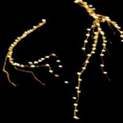
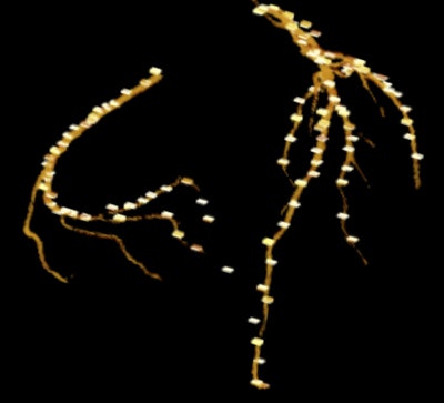
The results showed that vessels marked by MSCAR overlapped with 83.6% (8,520/10,191) of the center points marked manually by the radiologists. In addition, when compared with the 865 segments marked by radiologists, sensitivity reached 91.9% if a true positive was defined as a computer-segmented vessel that overlapped with at least 10% of the reference center points on a given segment. Increasing the threshold to 50% cut sensitivity to 86.2%, and increasing it to 100% left a sensitivity of just 53.4%.
 Radiologist-marked center points are superimposed on computer-extracted vessels rendered in 3D volume. Image courtesy of Chuan Zhou, PhD.
The group found a total of 55 false positives in 23 of the 62 test cases. MSCAR delivered high sensitivity for coronary artery segmentation and tracking, the authors concluded.
"In our study, the radiologists visually looked at each case and manually identified each false positive," Zhou said. "We are the first to count false positives, and there was less than one false positive per case."
A number of complementary studies are underway, he said. This includes efforts to improve MSCAR's accuracy for arterial segments affected by motion artifacts, severe calcifications, and noncalcified soft plaques, as well as to reduce the tracking of veins and noisy structures that are not coronary arteries. Research on flow tracking is also being conducted to determine the significance of detected stenosis.
The CAD side of the project
The accuracy of the tracking and segmentation scheme plays an important role in supporting what could be called the other half of the project, a CAD scheme that is detailed in another paper from the radiology department in the same issue of Medical Physics. The CAD scheme aimed to detect soft plaques, which is also an interesting project because soft plaques are far harder to see than calcium in the arteries, as their CT values are similar to those of soft tissues, Zhou said.
"Going from here, the continuous work is to detect the soft plaque," Zhou said. Using the CAD scheme developed to work on this segmentation method, "right now sensitivity is about 80% and false positives are about three to five per case."
But more than attenuation is involved in soft-plaque detection.
"We look at the CT values and we look at the shape of the changes around the vessel," he said. "The CAD does that using a feature set. We want to detect it and also measure the size of the occlusion by soft plaque."


