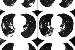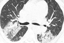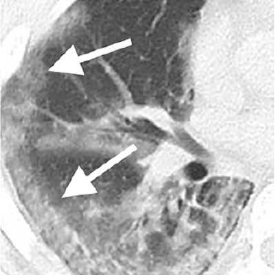
What should radiologists look for in cases suspicious for the novel coronavirus (2019-nCoV)? Researchers from the U.S. and China discussed the quintessential CT features of the virus that can aid in its early detection and diagnosis in a special report published online February 4 in Radiology.
The number of suspected 2019-nCoV cases worldwide has already surpassed 20,000, with at least 400 deaths, according to recent reports. A preliminary investigation into the first 41 confirmed 2019-nCoV cases showed that the patients' CT scans uniformly showed bilateral abnormal lung opacities.
Expanding upon this work, the authors of the current study examined the imaging data of 21 different patients infected by 2019-nCoV in an effort to characterize the most prevalent findings for radiologists as they help clinical teams address the outbreak.
"Early disease recognition is important not only for prompt implementation of treatment but also for patient isolation and effective public health surveillance, containment, and response," noted lead author Dr. Michael Chung, from the Icahn School of Medicine at Mount Sinai in New York, in a statement.
The new paper covers patients who were admitted to hospitals in China and underwent at least one chest CT exam. Their average age was 51.2 years, and all were confirmed positive for 2019-nCoV based on laboratory testing.
After analyzing the patients' imaging data, Chung and colleagues recognized that the most common CT findings were bilateral ground-glass opacities and consolidative pulmonary opacities, which were present on the scans of all patients with an abnormal CT scan. The only patients whose scans did not show one of these two types of opacities were the three patients who had entirely normal CT scans at presentation.
Three secondary CT findings helpful for early diagnosis were nodular opacities (33% of patients), a peripheral distribution of disease throughout the lungs (21%), and a crazy-paving pattern (19%).
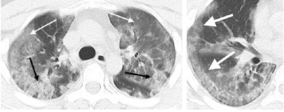 CT scans of a 29-year-old male with the novel coronavirus showing diffuse bilateral confluent and patchy ground-glass opacities and consolidative opacities (left). A zoomed-in look of the right middle and lower lobes shows striking peripheral distribution (right). Image courtesy of the RSNA.
In addition, there was no evidence of lung cavitation, discrete pulmonary nodules, pleural effusions, or lymphadenopathy in cases of 2019-nCoV. Follow-up CT scans demonstrated mild or moderate disease progression, as indicated by increasing extent and density of lung opacities, in roughly 88% of the patients.
A troubling aspect of the coronavirus is that infected patients can present with completely normal CT scans due to the long and variable incubation period of 2019-nCoV, the authors noted. In one case, a patient had normal chest CT results not only upon initial examination but also on a follow-up CT exam four days later, suggesting that clinicians cannot rely on CT alone to fully exclude the virus.
Not surprisingly, the overall imaging findings for 2019-nCoV resemble those reported for severe acute respiratory syndrome (SARS) and Middle East respiratory syndrome (MERS), both of which are also caused by coronaviruses, stated Dr. Jeffrey Kanne, from the University of Wisconsin-Madison School of Medicine, in an accompanying editorial.
"As the number of reported cases of 2019-nCoV infection continues to increase, radiologists may encounter [increasingly more] patients with this infection," Kanne wrote. Though detailed exposure and travel history are most critical to diagnosing 2019-nCoV, "bilateral ground-glass opacities or consolidation at chest imaging should prompt the radiologist to suggest 2019-nCoV as a possible diagnosis."

