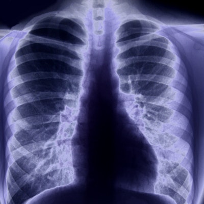
X-ray may not be the best imaging tool for detecting novel coronavirus disease (COVID-19). Almost three-quarters of a small cohort of South Korean patients with COVID-19 pneumonia had normal chest x-rays, missing pulmonary nodules that chest CT identified, according to a February 26 study in the Korean Journal of Radiology.
Although chest x-ray has been effective in diagnosing other coronaviruses, such as severe acute respiratory syndrome (SARS) and Middle East respiratory syndrome (MERS), radiography doesn't seem to be as effective with COVID-19. The new findings further support the argument for using CT to confirm COVID-19 infection, wrote a team led by Dr. Soon Ho Yoon, PhD, of Seoul National University Hospital.
"The proportion of patients with abnormal initial radiographic findings was 78.3% to 82.4% in SARS and 83.6% in MERS, but only 33% in our cases of COVID-19 pneumonia," the authors noted.
The outbreak of the COVID-19 virus has become a global emergency, on par with the outbreaks of SARS in 2003 and MERS in 2012. The first patient with COVID-19 in South Korea was identified on January 20, according to the researchers. The latest numbers from the World Health Organization as of March 2 indicate that there are 4,212 cases in the country, with 22 fatalities.
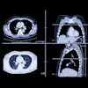
For the study, Yoon's group assessed image data from nine patients infected with COVID-19 who underwent chest x-ray and CT scans. Two radiologists interpreted the exams, checking whether abnormal findings on x-ray corresponded with those on CT.
They found that three of the nine patients (33%) had parenchymal abnormalities on chest x-ray, and most of these were peripheral consolidations. However, chest CT images showed double lung involvement in eight of the nine patients. In fact, a total of 77 pulmonary lesions were identified on chest CT in the patient cohort; of these, 39% were patchy lesions, 13% were large confluent lesions, and 48% were small nodular lesions. In 78% of these CT-identified lesions, peripheral lung fields were involved, while in 67%, posterior lung fields were involved.
Although the study highlights x-ray's limitations in diagnosing COVID-19, the authors did acknowledge that their patient cohort size was one of the research's limitations.
"The included patients only accounted for approximately one-third of all 29 identified patients of COVID-19 in Korea as of February 16, 2020," the group wrote. "Including more patients would enable a more comprehensive description of radiologic findings of COVID-19. However, we weighed this consideration against the importance of urgent reporting."
Since most of the pulmonary lesions identified on chest CT weren't clear on chest x-ray, it's important that radiologists be up to date on what to look for on CT, according to the team.
"Clinicians and radiologists should become familiar with the CT findings of COVID-19 and the limitations of chest radiographs in evaluating pneumonia to manage the COVID-19 outbreak," the group concluded.


.png?auto=format%2Ccompress&fit=crop&h=167&q=70&w=250)