Sunday, November 27 | 9:00 a.m.-10:00 a.m. | S1-SSCH01-5 | Room E351
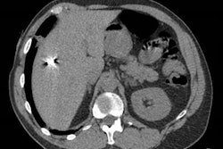
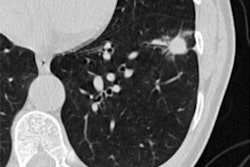
The effects of COVID-19 on patients' lungs are evident on CT, even a year after initial infection, according to this Sunday morning presentation.Presenter Dr. Martine Remy-Jardin, PhD, of the University Centre of Lille in France will share results from a study that included 79 patients who were hospitalized for COVID-19 pneumonia between March 2020 and April 2021. Patients were followed-up between six and 12 months with a dual-energy CT angiography exam and review of CT lung perfusion images.
COVID-19 patients continued to show abnormalities in their lungs in the year following hospitalization, the team found. CT showed acute pulmonary embolism features in two patients (2.5%) and focal chronic thromboembolic disease in three (3.8%), it also showed residual post-COVID-19 lung infiltration involving 4.7% of lung volume in 85% of patients. Radiologist readers rated lung perfusion as abnormal in 87.4% of study participants: These abnormal findings included patchy defects, areas of nonsystematized hypoperfusion, and pulmonary embolism-like defects.
The findings could give clinicians a better understanding about how COVID-19 affects people long-term.
"[Later] follow-up showed CT features of acute and chronic pulmonary embolism but also two types of perfusion abnormalities suggestive of persistent hypercoagulability as well as unresolved sequelae of the widespread [cerebral small vessel disease] described in the acute phase of the disease," the group concluded.


