CHICAGO -- A fully automated AI algorithm designed to opportunistically screen for enlarged heart, or cardiomegaly, on routine chest and abdominal CT exams was highly predictive of future cardiovascular events, according to research presented November 26 at RSNA 2023.
Presenter Steven Rothenberg, MD, of the University of Alabama in Birmingham, said calculating the cardiothoracic ratio (CTR) was most useful for opportunistic screening of cardiomegaly on cross-sectional imaging.
“We hypothesized that a suite of fully automated algorithms that quantifies the cardiothoracic ratio would predict future cardiovascular events,” Rothenberg explained. “The reason why this is important is because early detection of cardiomegaly would allow for appropriate management to reduce cardiovascular events. And cardiovascular disease is the number one cause of mortality in the U.S.”
Cardiomegaly is pathologic enlargement of the heart that can be caused by chronic conditions. Having cardiomegaly predisposes a person to major cardiovascular events such as myocardial infarction, heart failure, stroke, arrhythmia, or death.
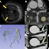
For this study, Rothenberg's group trained the AI algorithm to segment the heart, determine the key slice for where the heart is largest, and then extract the cardiothoracic ratio from 2D segmentation. A normal heart was defined as having a linear cardiothoracic ratio of less than 0.5, while mild cardiomegaly was defined as a ratio of 0.5 to 0.56, and severe cardiomegaly as a ratio greater than 0.56. The team gathered these thresholds from exploratory statistics and a background literature search.
"We created an objective way to automate the measurement of heart size," Rothenberg said in an interview with AuntMinnie.com.
When asked why CTR was preferred over chamber volume, Rothenberg said that while chamber volume might be more specific, it would be more difficult to get accurate results in noncontrast exams. Chamber volume would also be inaccurate on abdominal CT scans because the entire heart might not be included in the field of view. Instead, CTR is reproducible, broadly generalizable, and easy for radiologists to verify AI outputs or calculate manually. He added that three board-certified radiologists validated the model.
This opportunistic screening study was limited with its single-center retrospective approach. However, a very large sample size was used (14,299), and researchers also had ample time (six years) to collect cardiovascular events that occurred with those in the study group, Rothenberg said. An i2b2 search of ICD codes identified cardiovascular events. A search of electronic medical records helped classify patients as unmanaged or managed for cardiomegaly (e.g., echocardiogram in the prior year). Single-variable Cox proportional hazards regression models were used to associate the CTR with future cardiovascular events.
Using the algorithm, researchers were able to classify 8,643 as normal, 4,188 as mild cardiomegaly, and 1,468 with severe cardiomegaly. In addition, Rothenberg said a time-to-event analysis showed patients with severe cardiomegaly were 4.2 times more likely to have a myocardial infarction; 3.6 times more likely to have heart failure; 2.2 times more likely to have stroke; two times more likely to have an arrhythmia; two times more likely to have any cardiovascular event; and 1.8 times more likely to die.
When the initial version of the algorithm did not perform well on difficult cases, Rothenberg said the team enriched the training set with pathologies such as pleural effusion, pneumonia, and pneumonectomy to be more representative of the distribution of cases encountered in daily practice that might challenge the algorithm. He said the model applies to routine chest and abdominal CT with or without contrast and could be used for opportunistic screening for cardiomegaly.
“We didn't do a large-scale validation on multi-institutional data,” Rothenberg said. “I think that what would be an interesting next step for this research. The data we get on our CT scans is a goldmine of information about someone’s overall health. Adding this data gives us an opportunity to identify patients who are at risk and make sure they get appropriate care downstream."