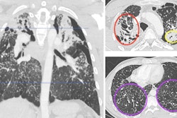
Interobserver agreement for assessing interstitial lung disease (ILD) using high-resolution CT (HRCT) is moderate, researchers have reported.
The findings highlight "current weaknesses in knowledge and diagnostic criteria and should help radiologists realize the limitations of HRCT and stress the importance of multidisciplinary team discussion and other clinical parameters in patient care," wrote a team led by Liam Delaney, MBChB, of the University of Sheffield Royal Hallamshire Hospital in the U.K. The study was published October 15 in Radiology.
"Our results demonstrate that repeatable assessment of disease severity, extent, and progression is challenging even for expert radiologists," the group noted.
High-resolution CT is a key imaging modality for evaluating ILD, the investigators explained, noting that "accurate classification of disease has important implications for patients." But assessing imaging features for the condition can be challenging, even for experienced thoracic radiologists.
Since previous studies have produced mixed results regarding the interpretation of high-resolution CT features on interstitial lung disease-related imaging, Delaney and colleagues conducted an analysis of 13 studies published between January 2000 and October 2023 on the Ovid MEDLINE, Embase, and Cochrane Central Register of Controlled Trials databases to determine level of agreement on CT imaging of ILD; they tracked the severity and progression of disease and the presence of features such as honeycombing and ground-glass opacification based on the 2011 and 2018 American Thoracic Society/European Respiratory Society/Japanese Respiratory Society/Asociación Latinoamericana del Tórax (ATS/ERS/JRS/ALAT) guidelines for idiopathic pulmonary fibrosis (IPF). The 13 studies included 6,943 images and 146 radiologists. The authors measured interobserver agreement using pooled κ or intraclass correlation coefficient (ICC) values.
Overall, the investigators only found modest interobserver agreement:
- Among 10 studies that evaluated agreement of specific radiologic findings in ILD, the pooled κ value was 0.56 (with 1 as reference).
- Among eight studies the interobserver agreement of the ATS/ERS/JRS/ALAT diagnostic guidelines for IPF based on typical UIP patterns, the pooled κ value was 0.61.
- One of the 13 studies found a pooled κ value of 0.87 for ILD progression.
- Seven studies that evaluated ILD severity could not be combined, the team reported: κ values for ILD severity varied from 0.64 to 0.90, and intraclass correlation coefficient values from 0.63 to 0.96.
The results underscore the need for more research, according to the group.
"The study highlights current weaknesses in knowledge and diagnostic criteria and should help radiologists realize the limitations of [high-resolution CT and stress the importance of multidisciplinary team discussion and other clinical parameters in patient care," it concluded.
The complete study can be found here.


