Using CT to track the distribution of lymph nodes in colon cancer patients is more accurate for identifying metastasis than conventional clinical stage assessment, according to a study published April 8 in Radiology.
This can be particularly important in predicting treatment response in patients with "microsatellite instability-high" (MSI-H) colon cancer, according to a team led by Zhen Guan, PhD, of Peking University Cancer Hospital and Institute in Beijing, China. (MSI-H is characterized by cancer cells manifesting a high number of mutations in short, repeated DNA sequences, i.e., microsatellites.)
"[We found that distribution-based clinical lymph node stage] showed high performance for identifying regional lymph node metastases and helped predict complete response after neoadjuvant immunotherapy in MSI-H colon cancer using a surgical reference standard," Guan and colleagues wrote.
Identifying lymph node metastasis is crucial for cases of MSI-H colon cancer, but CT's accuracy can be limited, the researchers explained. They investigated whether using CT to identify lymph node distribution patterns could improve lymph node evaluation in the disease via a study that included 368 patients who underwent pretreatment CT and surgery between December 2017 and January 2024. (Of the total cohort, 230 made up a development set [used to create a distribution-based clinical lymph node stage]; 86 made up a test set; and 52 made up a treatment set. The development set underwent CT and surgery between December 2017 to December 2022, while the test set underwent the exam and procedure between January 2016 and January 2024).
The investigators used lymph node characteristics associated with pathologic lymph node metastasis such as clinical lymph node stage and distribution patterns (vascular distribution, jammed cluster, and partial fusion) to create the distribution-based clinical lymph node stage algorithm in the development set. The group then assessed the development set's diagnostic performance in the test set and evaluated its clinical value in the treatment cohort (these patients underwent neoadjuvant immunotherapy between August 2017 and February 2024).
The researchers noted that only jammed cluster and partial fusion were associated with higher odds of pathologic lymph node metastasis (odds ratio, 78.9 and 21.5, respectively; both p< 0.001). They also found the following:
| Performance of distribution-based clinical lymph node stage vs. clinical lymph node stage for predicting colon cancer treatment response | ||
|---|---|---|
| Measure | Clinical lymph node stage | Distribution-based clinical lymph node stage |
| Accuracy | 46% | 90% |
| Specificity | 26% | 97% |
| Interobserver agreement (kappa value, with 1 as reference) | 0.48 | 0.67 |
Finally, the team reported that distribution-based clinical lymph node stage was associated with a complete response after neoadjuvant immunotherapy, with an odds ratio of 0.05 (p< 0.001).
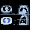
 Schematic diagrams and representative images of different distribution patterns of region lymph nodes detected at CT. (A) Vascular distribution pattern: All regional lymph nodes detected at CT have regular borders and are distributed along the vessels (arrowheads). (B-E) Coronal CT images in a (B) 34-year-old woman, (C) 51-year-old man, (D) 37-year-old man, and (E) 40-year-old man with microsatellite instability-high (MSI-H) colon cancer show multiple clear lymph nodes distributed along the supplying arteries or draining veins (arrowheads). (F) Jammed cluster pattern: At least three lymph nodes with blurred margins and clustered (circle). (G-I) Coronal CT images in a (G) 29-year-old man, (H) 35-year-old man, and (I) 67-year-old man and (J) axial CT image in a 43-year-old man with MSI-H colon cancer show several lymph nodes with blurred margins and clustered (circle). Movies 1-4 provide more details. (K) Fused pattern: Several poorly defined lymph nodes clustered and fused together (arrow). (L-O) Coronal CT images in a (L) 63-year-old woman, (M) 59-year-old man, (N) 49-year-old woman, and (O) 73-year-old man with MSI-H colon cancers show several lymph nodes fused (arrow). Image courtesy of Radiology.
In an accompanying commentary, Naama Lev-Cohain, MD, and colleague Jacob Sosna, MD, both of Hadassah Medical Center in Jerusalem, Israel, noted that the study "presents a compelling approach to improving lymph node staging in MSI-H colon cancer, offering potential benefits for tailored treatment and better patient outcomes."
"Unlike traditional lymph node evaluation methods, which focus on individual nodal characteristics, Guan and Wang et al propose a broader, pattern-based distribution approach," the two wrote. "This distribution-based staging method significantly improves diagnostic performance, surpassing the conventional approach."
The complete study can be found here.