
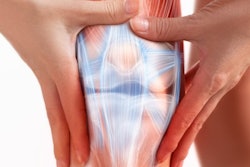
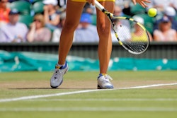
Lateral epicondylitis -- also known as tennis elbow -- can be effectively treated through an interventional radiology procedure called transcatheter arterial embolization (TAE), according to research presented at the Society of Interventional Radiology (SIR) annual meeting in Austin, TX.
The findings are good news for patients because lateral epicondylitis can be difficult to resolve, said lead author Dr. Yuji Okuno, PhD, of the Okuno Clinic in Tokyo, in a statement released by SIR.
"Tennis elbow can be difficult to treat, leaving many patients unable to perform the simplest tasks, such as picking up their children, cooking dinner, or even working on a computer," he said. "With this frustration, many patients turn to invasive major surgery after years of failed physical therapy and medication use. We were interested to see if this technique ... would be effective for this common, debilitating condition and help people immediately regain a range of motion that many of us take for granted."
Okuno and colleagues conducted a study that included 48 patients with lateral epicondylitis who underwent TAE between March 2013 and October 2017 and then were followed for four years. The investigators used the Quick Disabilities of the Arm, Shoulder, and Hand pain score questionnaire to track patients' results at baseline and at one, three, six, and 24 months post-treatment.
The group found statistically significant reductions in the scores, with an overall score of 52.1 at baseline and 3.7 at 24 months. In addition, on MRI scans performed two years after treatment, 32 of the 48 patients showed improvement in tendinosis and tear scores compared to baseline, and no patients showed bone marrow necrosis, cartilage loss, or muscle atrophy.
The TAE procedure takes about an hour, and it uses a catheter inserted at the radial artery in the wrist under local anesthesia, according to the investigators.
The study findings suggest that TAE is a feasible treatment option for patients with tennis elbow who do not respond to conservative treatments for the condition, they concluded.


![Representative example of a 16-year-old male patient with underlying X-linked adrenoleukodystrophy. (A, B) Paired anteroposterior (AP) chest radiograph and dual-energy x-ray absorptiometry (DXA) report shows lumbar spine (L1 through L4) areal bone mineral density (BMD). The DXA report was reformatted for anonymization and improved readability. The patient had low BMD (Z score ≤ −2.0). (C) Model (chest radiography [CXR]–BMD) output shows the predicted raw BMD and Z score in comparison with the DXA reference standard, together with interpretability analyses using Shapley additive explanations (SHAP) and gradient-weighted class activation maps. The patient was classified as having low BMD, consistent with the reference standard. AM = age-matched, DEXA = dual-energy x-ray absorptiometry, RM2 = room 2, SNUH = Seoul National University Hospital, YA = young adult.](https://img.auntminnie.com/mindful/smg/workspaces/default/uploads/2026/04/ai-children-bone-density.0snnf2EJjr.jpg?auto=format%2Ccompress&fit=crop&h=100&q=70&w=100)


![Representative example of a 16-year-old male patient with underlying X-linked adrenoleukodystrophy. (A, B) Paired anteroposterior (AP) chest radiograph and dual-energy x-ray absorptiometry (DXA) report shows lumbar spine (L1 through L4) areal bone mineral density (BMD). The DXA report was reformatted for anonymization and improved readability. The patient had low BMD (Z score ≤ −2.0). (C) Model (chest radiography [CXR]–BMD) output shows the predicted raw BMD and Z score in comparison with the DXA reference standard, together with interpretability analyses using Shapley additive explanations (SHAP) and gradient-weighted class activation maps. The patient was classified as having low BMD, consistent with the reference standard. AM = age-matched, DEXA = dual-energy x-ray absorptiometry, RM2 = room 2, SNUH = Seoul National University Hospital, YA = young adult.](https://img.auntminnie.com/mindful/smg/workspaces/default/uploads/2026/04/ai-children-bone-density.0snnf2EJjr.jpg?auto=format%2Ccompress&dpr=2&fit=crop&h=167&q=70&w=250)


