
Konica Minolta Healthcare Americas and Shimadzu Medical Systems USA are partnering to bring new dynamic digital radiography (DDR) technology to the U.S. market.
First demonstrated by Konica Minolta at RSNA 2018, DDR is a new technology that enables the visualization of movement in conventional x-ray exams without fluoroscopy. DDR is an enhanced version of a standard DR system designed to acquire up to 15 sequential radiographs per second for up to 20 seconds of physiological movement. The result is 300 x-ray images acquired with a dose equivalent to approximately two standard x-rays.
DDR may be promising for use in thoracic imaging because it allows clinicians to observe chest wall, heart, and lung motion during respiration. It could also be used for orthopedic applications in the spine and extremities.
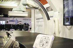
Konica Minolta and Shimadzu collaborated in the development of DDR by combining Konica Minolta's image processing technologies and Shimadzu's RADspeed Pro radiography system. Under the partnership signed between the firms, both companies will co-market DDR technology in the U.S.
The firms will show DDR on RADspeed Pro at the AHRA annual meeting this week in Denver.


![Representative example of a 16-year-old male patient with underlying X-linked adrenoleukodystrophy. (A, B) Paired anteroposterior (AP) chest radiograph and dual-energy x-ray absorptiometry (DXA) report shows lumbar spine (L1 through L4) areal bone mineral density (BMD). The DXA report was reformatted for anonymization and improved readability. The patient had low BMD (Z score ≤ −2.0). (C) Model (chest radiography [CXR]–BMD) output shows the predicted raw BMD and Z score in comparison with the DXA reference standard, together with interpretability analyses using Shapley additive explanations (SHAP) and gradient-weighted class activation maps. The patient was classified as having low BMD, consistent with the reference standard. AM = age-matched, DEXA = dual-energy x-ray absorptiometry, RM2 = room 2, SNUH = Seoul National University Hospital, YA = young adult.](https://img.auntminnie.com/mindful/smg/workspaces/default/uploads/2026/04/ai-children-bone-density.0snnf2EJjr.jpg?auto=format%2Ccompress&dpr=2&fit=crop&h=167&q=70&w=250)


