

Monday, November 29 | 1:30 p.m.-2:30 p.m. | SSPH04-5 | Room TBA
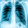
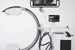
In this talk, Ran Zhang, PhD, will present a prototype developed by a group of medical physicists at the University of Wisconsin-Madison of a low-dose, multicontrast chest x-ray system that can provide three mutually complementary images from a single acquisition. The new system is fast and eventually could be put to use diagnosing and screening for lung cancer, the team suggests.The researchers built a multicontrast chest x-ray system that can provide conventional absorption contrast, differential phase contrast, and dark-field x-ray contrast images in a single acquisition. They implemented several technical innovations to achieve data acquisition of the entire chest with a coverage of 28 cm along the superior-inferior direction within four seconds. Initial imaging tests were performed using a chest phantom.
The estimated air kerma and effective dose were well below the effective dose for a typical chest x-ray and the quality of the absorption contrast images matched conventional chest x-rays, the group found.
"A new prototype multicontrast [chest x-ray] system was developed to enable low radiation dose and fast scans of human chest-sized objects within four seconds," Zhang and colleagues noted.
Attend this session to learn the details.
This paper received a Roadie 2021 award for the most popular abstract by page views in this Road to RSNA section.
![Representative example of a 16-year-old male patient with underlying X-linked adrenoleukodystrophy. (A, B) Paired anteroposterior (AP) chest radiograph and dual-energy x-ray absorptiometry (DXA) report shows lumbar spine (L1 through L4) areal bone mineral density (BMD). The DXA report was reformatted for anonymization and improved readability. The patient had low BMD (Z score ≤ −2.0). (C) Model (chest radiography [CXR]–BMD) output shows the predicted raw BMD and Z score in comparison with the DXA reference standard, together with interpretability analyses using Shapley additive explanations (SHAP) and gradient-weighted class activation maps. The patient was classified as having low BMD, consistent with the reference standard. AM = age-matched, DEXA = dual-energy x-ray absorptiometry, RM2 = room 2, SNUH = Seoul National University Hospital, YA = young adult.](https://img.auntminnie.com/mindful/smg/workspaces/default/uploads/2026/04/ai-children-bone-density.0snnf2EJjr.jpg?auto=format%2Ccompress&fit=crop&h=100&q=70&w=100)


![Representative example of a 16-year-old male patient with underlying X-linked adrenoleukodystrophy. (A, B) Paired anteroposterior (AP) chest radiograph and dual-energy x-ray absorptiometry (DXA) report shows lumbar spine (L1 through L4) areal bone mineral density (BMD). The DXA report was reformatted for anonymization and improved readability. The patient had low BMD (Z score ≤ −2.0). (C) Model (chest radiography [CXR]–BMD) output shows the predicted raw BMD and Z score in comparison with the DXA reference standard, together with interpretability analyses using Shapley additive explanations (SHAP) and gradient-weighted class activation maps. The patient was classified as having low BMD, consistent with the reference standard. AM = age-matched, DEXA = dual-energy x-ray absorptiometry, RM2 = room 2, SNUH = Seoul National University Hospital, YA = young adult.](https://img.auntminnie.com/mindful/smg/workspaces/default/uploads/2026/04/ai-children-bone-density.0snnf2EJjr.jpg?auto=format%2Ccompress&dpr=2&fit=crop&h=167&q=70&w=250)


