Virtual Phantoms has launched VirtualDoseDX, a radiation dose estimator for diagnostic x-ray.
VirtualDoseDX allows users to adjust x-ray output and orientation to match site-specific techniques or patient protocols. It can be used by radiology teams to standardize patient dose on x-ray exams, by researchers to explore population radiation exposure, or by institutions seeking to meet regulatory and accreditation standards, according to the firm.
The company has also developed VirtualDoseCT for CT imaging and VirtualDoseIR for interventional radiology dose estimation, it said. VirtualDoseDX is available as a standalone offering or as part of the firm's VirtualDose product suite.


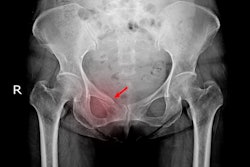


![Representative example of a 16-year-old male patient with underlying X-linked adrenoleukodystrophy. (A, B) Paired anteroposterior (AP) chest radiograph and dual-energy x-ray absorptiometry (DXA) report shows lumbar spine (L1 through L4) areal bone mineral density (BMD). The DXA report was reformatted for anonymization and improved readability. The patient had low BMD (Z score ≤ −2.0). (C) Model (chest radiography [CXR]–BMD) output shows the predicted raw BMD and Z score in comparison with the DXA reference standard, together with interpretability analyses using Shapley additive explanations (SHAP) and gradient-weighted class activation maps. The patient was classified as having low BMD, consistent with the reference standard. AM = age-matched, DEXA = dual-energy x-ray absorptiometry, RM2 = room 2, SNUH = Seoul National University Hospital, YA = young adult.](https://img.auntminnie.com/mindful/smg/workspaces/default/uploads/2026/04/ai-children-bone-density.0snnf2EJjr.jpg?auto=format%2Ccompress&dpr=2&fit=crop&h=167&q=70&w=250)

