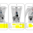
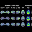
PHILADELPHIA - Although FDG-PET has shown a high degree of accuracy in discriminating between normal patients and those with Alzheimer's disease (AD), not much is known about the modality's ability to differentiate between AD and frontotemporal dementia (FTD). A group of researchers from Belgium, Germany, and Italy presented the results of a prospective multicenter study that sought to determine the potential of PET in discriminating between AD and FTD.
Dr. Karl Herholz of the Max-Planck-Institute for Neurological Research at the University of Cologne, in Germany, presented this study Monday at the Society of Nuclear Medicine meeting. The study was supported by the European Network for Efficiency and Standardization of Dementia Diagnosis (NEST-DD), a multicenter task force working to improve and standardize the diagnosis of dementia.
For this study, the investigators processed each scan with spatial normalization and subtraction of projections. The resulting images were then examined against the principal components of a multivariate 3D voxel-based map of brain functional imaging data, which represented the normal variability of FDG-PET scans across the centers.
The correlation of these images with average AD and FTD images was determined and subjected to discriminant analysis, Herholz said. The resulting linear discriminant function achieved an 89% correct assignment in a retrospective sample of 544 subjects (394 AD, 40 FTD, 110 controls).
"This discriminant function was applied to a separate prospective sample of 246 patients, with 214 probable Alzheimer's patients and 32 FTD patients," he said.
The correspondence between PET and clinical classification in the prospective sample was 85%, according to the researchers. Correspondence was better in clinical AD (89%) than in clinical FTD, from which group only 18 of 32 patients were classified by PET as FTD, but 14 as AD.
Comparison with clinical symptoms showed that FDG-PET classification as FTD was more closely related to impairment of language fluency than to apathy and disinhibition.
"In many cases, overlap of AD and FTD pattern features was present," noted Herholz.
The group found that the PET-based discriminant function provided a significant classification (at p < 0.05) in 91 cases, of which 90% corresponded with the clinical classification (91% in AD, 84% in FTD). Visual inspection of the PET images confirmed the classification by the discriminant function, although Herholz noted that the team had less certainty of discrimination at a higher age of onset of dementia.
"The margin of FDG-PET to improve clinical discrimination between AD and FTD at the clinical stage of mild dementia is in the order of 10%-15% of cases," he said.
Based on the results, Herholz said the researchers were contemplating further studies to address the interaction of frontal hypometabolism with aging and vascular change, the use of improved image-analysis techniques, and an assessment of early diagnosis and long-term follow-up using autopsy-proven diagnoses.
By Jonathan S. BatchelorAuntMinnie.com staff writer
June 23, 2004
Related Reading
CMS to reimburse PET for Alzheimer's, June 16, 2004
Using PET for Alzheimer's diagnosis lowers cost of care, October 21, 2002
FDG-PET depicts metabolic patterns of Alzheimer's in young adults, July 24, 2002
U.S. gives thumbs-down on PET for Alzheimer's, January 1, 2002
PET, SPECT differ in Alzheimer's imaging, June 25, 2001
Copyright © 2004 AuntMinnie.com


