PET/CT imaging with an experimental gallium-68-based (Ga-68) radiotracer that binds to overactive fibroblast cells in tumors outperforms standard methods for detecting the spread of lung cancer, according to a study published January 4 in Radiology.
In a prospective study in patients with advanced lung cancer, Chinese researchers compared PET/CT imaging using a Ga-68-labeled fibroblast-activation protein inhibitor (FAPI) radiotracer versus standard F-18 FDG imaging for detecting lung cancer metastases. They found that Ga-68 FAPI PET/CT and F-18 FDG PET/CT performed similarly in detecting primary tumors and metastases in the lungs but that Ga-68 FAPI edged ahead when detecting brain and bone metastases.
"[Ga-68 FAPI] PET/CT may outperform [F-18 FDG] PET/CT in staging lung cancer, particularly in the detection of metastasis to the brain, lymph nodes, bone, and pleura," wrote co-first authors Dr. Lijuan Wang and Ganghua Tang, PhD, of the Nanfang PET Center at Southern Medical University in Guangzhou.
Brain and bone metastases are a major cause of death in patients with lung cancer. Current guidelines recommend pretreatment F-18 FDG PET/CT scans combined with contrast-enhanced brain MRI to assist in clinically staging patients, yet high background radioactivity in the brain seen in F-18 FDG PET/CT results in low sensitivity for detecting brain metastasis, according to the authors.
The observation that fibroblast-activation protein is overexpressed in cancerous fibroblast cells in tumors has led to the recent development of experimental PET tracers that combine FAP inhibitors with Ga-68. These tracers have demonstrated promising results preclinically for detecting numerous types of cancer and are currently being investigated in trials.
In this study, the authors investigated whether Ga-68 FAPI PET/CT is superior to imaging with F-18 FDG PET/CT for detecting lung cancer, particularly of metastasis to the brain, lymph nodes, bone, and pleura.
The researchers enrolled 34 advanced lung cancer patients between the ages of 46 and 80 from September 2020 to February 2021 at Southern Medical University's Nanfang Hospital. They included participants with newly diagnosed stage IV lung cancer who had not received any prior treatment or patients with recurrent or post-treatment metastatic lung cancer who had not received any treatment for more than three months. Patients underwent both Ga-68 FAPI and F-18 FDG PET/CT scans (uExplorer, United Imaging) within one week of each other.
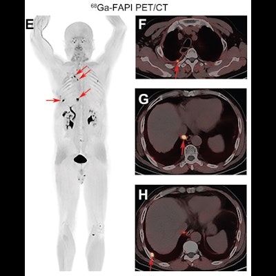
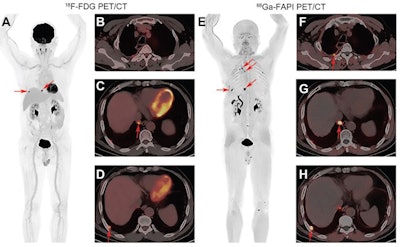
 Images of a 56-year-old man with lung adenocarcinoma for tumor restaging after treatment. (A) Maximum intensity projection F-18 FDG image, and (B-D) axial fused F-18 FDG PET/CT images of upper chest and upper abdomen. (E) Maximum intensity projection Ga-68 FAPI image and (F-H) axial fused Ga-68 FAPI PET/CT images of upper chest and upper abdomen. Ga-68 FAPI PET/CT images depict four positive lesions (arrows) in the right pleura with intense tracer uptake (SUVmax: 4.9 to approximately 12.8). F-18 FDG PET/CT images only depict two positive lesions in the right pleura with a slightly lower tracer uptake (arrows in A, C, and D; SUVmax: 8.5-10.4). Image courtesy of Radiology.
F-18 FDG PET/CT images were reviewed independently by two nuclear oncologists with 13 and 15 years of board-certified experience, and Ga-68 FAPI PET/CT images were reviewed by two nuclear oncologists with 18 and 27 years of experience. Disagreements were resolved by consensus.
The study authors found both tracers showed similar performance in the delineation of primary tumors and detection of suspected metastases in the lungs, liver, and adrenal glands. The metabolic tumor volume in primary and recurrent lung tumors showed no differences between Ga-68 FAPI and f_18 FDG (11.6 versus 10.8).
However, compared with F-18 FDG PET/CT, Ga-68 FAPI PET/CT revealed more suspected metastases in lymph nodes (356 vs. 320), brain (23 vs. 10), bone (109 vs. 91), and pleura (66 v.s 35), the authors stated.
Semiquantitative evaluation showed the SUVmax and target-to-background ratios (TBR) of primary or recurrent tumors, positive lymph nodes, bone lesions, and pleural lesions on Ga-68 FAPI PET/CT were all higher than those on F-18 FDG PET/CT (all, p < 0.01).
In addition, although SUVmax of Ga-68 FAPI and F-18 FDG in brain metastases were not different (mean SUVmax: 9 vs. 7.4, p = 0.32), TBR was higher with Ga-68 FAPI than with F-18 FDG (mean: 314.4 vs. 1, p = 0.02), the researchers stated.
"[Ga-68 FAPI PET/CT] has the potential to be an alternative to F-18 FDG for clinical staging examinations," they wrote.
In an accompanying editorial, Drs. Francine Jacobson and Annick Van den Abbeele of Harvard Medical School noted that at least 28 different cancers so far have demonstrated Ga-68 FAPI avidity. Further studies are required to validate the tracer.
The approach used by the authors of this study provides an efficient model for comparing the clinical utility of novel tracers that can be applied to other cancers besides lung cancer, and it may also open the door to novel diagnostic and therapeutic approaches, Jacobson and Abbeele wrote.
"Having a molecular imaging tracer that targets the tumor microenvironment may add enormous value to the characterization of cancer and complementary information to other molecular imaging tracers currently used in the clinic, including F-18 FDG," they concluded.


