PET/CT imaging shows that inflammation of the aorta is increased in the early postinfection stage of COVID-19 in patients with severe disease, according to a study published May 2 in the Journal of Nuclear Cardiology.
Greek researchers performed F-18 FDG-PET/CT scans in patients admitted with severe or critical COVID-19 infections at two local hospitals in Athens. They found the scans revealed significant inflammation in heart vessels in patients early after infection, and that the inflammation largely resolved over time.
"Our findings may have important implications for the understanding of the course of the disease and for improving our preventive and therapeutic strategies," wrote corresponding author Dr. Charalambos Vlachopoulos, a cardiologist at the University of Athens.
F-18 FDG-PET/CT imaging is a valuable tool for the diagnosis and assessment of disease severity in different types of vascular and cardiac inflammation and infection, based on the metabolic activity of the F-18 FDG radiotracer revealed in the scans.
To date, studies are limited on aortic F-18 FDG uptake in patients infected with COVID-19, however, with one retrospective analysis in patients with long COVID-19 symptoms reporting a small number of patients had increased standard uptake values (SUVmax) in the thoracic aorta.
"This is the first study that investigates prospectively the presence of aortic inflammation during early recovery in COVID-19 in severely or critically ill patients," the authors wrote.
Vlachopoulos and colleagues recruited 20 patients (mean age of 59 ± 12 years) hospitalized with severe or critical COVID-19 between November 2020 and May 2021. Patients had a prior history of malignancy, but they were free of active disease at the time of the study.
Patients underwent whole-body PET/CT imaging between 20 and 120 days after hospital admission. Ten age and sex-matched individuals without COVID-19 who were scheduled for F-18 FDG-PET/CT imaging served as the control group.
Aortic inflammation was assessed by measuring 18F-FDG uptake in PET/CT performed 20-120 days after admission. Ten age and sex-matched individuals served as the control group. Global aortic target-to-background ratios (GLA-TBR) and index aortic segment TBR (IAS-TBR) were calculated to measure molecular inflammation activity.
The researchers observed no significant global differences in aortic F-18 FDG PET/CT uptake between COVID-19 patients and controls, yet patients who were scanned less than 60 days from admission (n = 11) had significantly higher values (GLA-TBR, 1.53) compared with patients scanned more than 60 days after admission (GLA-TBR, 1.40).
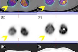
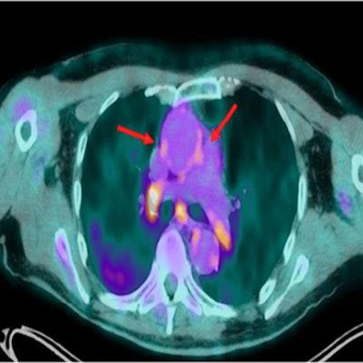
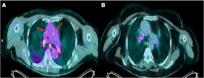 Transaxial views of fused F-18 FDG PET/CT images of two patients with COVID-19. (A) A patient scanned 20 days after admission with severe COVID-19. (B) A patient scanned 64 days after admission. In the former, there is increased F-18 FDG uptake in the wall of the ascending aorta (arrows), as well as hypermetabolic hilar and mediastinal lymph nodes. Image courtesy of the Journal of Nuclear Cardiology.
There was also a significant difference in IAS-TBR between patients scanned in less than 60 days (1.64) and controls (1.5), according to the findings.
"Aortic inflammation, as assessed by F-18 FDG PET/CT imaging, is increased in the early post-COVID phase in patients with severe or critical COVID-19 and largely resolves over time," Vlachopoulos et al wrote.
The findings may have important implications for the understanding of the course of the disease and for improving preventive and therapeutic strategies, the authors wrote.
"F-18 FDG-PET/CT imaging could recognize early patients that have residual aortic inflammation and may serve as a predictor of outcome both in the early and late postinfection period," they stated.
However, additional studies are required, the authors noted. This study is limited by its observational nature, as no individual patient serial measurements with subsequent F-18 FDG PET/CT scans were taken, they wrote.
"Further research is needed to disentangle the process of recovery of inflammation (aortic and systemic) in patients with COVID-19," Vlachopoulos and colleagues concluded.


