Dynamic PET scans acquired in half the time of normal scans appear clinically feasible for patients with lung lesions, according to nuclear medicine researchers in Shenzhen, China.
In an image reconstruction analysis, the group found that dynamic F-18 FDG-PET images acquired in 30 minutes provided images equal in quality to scans acquired after 65 minutes – a big difference for patients who are required to lie prone during the procedures, noted lead authors Fen Du, PhD, and Xieraili Wumener, PhD, of the Chinese Academy of Medical Sciences.
“Protocols with a shortened 30-minute acquisition time may be considered for patients who have difficulty with prolonged acquisitions,” the group wrote. The article was published March 5 in EJNMMI Physics.
F-18 FDG-PET scans may be static or dynamic, with static imaging capturing the distribution of radiotracer 60 minutes after it is injected in patients based on standard uptake values (SUVs), the authors explained. While more common, the technique may be limited in identifying malignant lung lesions versus benign (e.g., inflammatory) F-18 FDG tracer uptake, the authors noted.
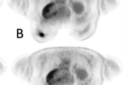
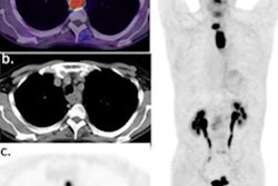
 A 60-year-old male patient with clinical suspicion of lung cancer. PET/CT scan showed FDG-avid pulmonary nodules in the upper lobe of the left lung (red arrow), with a size of 2.2 × 1.3 cm, SUVmax of 12.2, Ki-65 min of 0.016 ml/g/min, and Ki-30 min of 0.014 ml/g/min. Surgical pathology confirmed a granulomatous lesion. In addition, PET/CT scan showed many FDG-avid lung nodules in the mediastinum. (A, static PET maximum intensity projection; B, static PET SUV image; C, static PET/CT fusion image; D, dynamic PET Ki-65 min image; E, dynamic PET Ki-30 min image). Image courtesy of EJNMMI Physics.
Conversely, dynamic PET imaging is performed continuously over longer scan times, yet allows clinicians to measure what are called “kinetic rate constants” – parameters of radiotracer activity in lesions over time – and have proven to be more accurate than SUV measurements, the researchers added.
However, longer scan times of may not be suitable for certain patients, such as those with anxiety or mobility issues, the authors noted.
In this study, the researchers tested the clinical feasibility of early 30-minute dynamic PET in terms of image quality and diagnostic efficacy in benign and malignant lung lesions. The group analyzed dynamic F-18 FDG-PET images of 146 patients with 181 lung lesions (including 146 lesions confirmed by histology).
Using Matlab software, the researchers reconstructed the dynamic scans into 28 frames that showed kinetic rates (Ki) of F-18 FDG radiotracer uptake by the lung lesions over either 30 minutes or 65 minutes.
Based on a visual analysis by two experts, the quality of the Ki 30-minute images was not significantly different from the Ki 65-minute images, according to the authors. Moreover, an analysis of 146 cases of histologically confirmed lesions resulted in scores of 82% when differentiating lung lesions using both the Ki 65-minute and Ki 30-minute scans, the researchers added.
“In terms of image quality, our study found that the quality of Ki-30 min images is as good as that of Ki-65 min images by visual quality assessment,” the group wrote.
Ultimately, the main hurdle for dynamic FDG-PET imaging in achieving more clinical use is its longer scanning time and optimal acquisition times for various applications have yet to be determined, the authors noted.
“This study indicates that an early 30-minute dynamic FDG-PET acquisition appears to be sufficient to provide quantitative images with good-quality and accurate Ki values for the assessment of lung lesions,” the group concluded.
The full article can be found here.


![A 53-year-old patient (patient number four) with a recurrent pituitary adenoma with extension of a cystic component of disease to the medial temporal lobe apparent on MRI (contoured in blue), and extension of disease to the left sphenoid bone and orbital apex apparent on [68Ga]Ga-DOTA-TATE (contoured in yellow).](https://img.auntminnie.com/mindful/smg/workspaces/default/uploads/2026/04/pituitary-tumor.QGsEnyB4bU.jpg?auto=format%2Ccompress&dpr=2&fit=crop&h=167&q=70&w=250)


