A recent head-to-head study suggests that PET/CT imaging with a fibroblast activation protein inhibitors (FAPI) radiotracer is superior to F-18 FDG-PET/CT for assessing patients with liver cancer.
Moreover, the approach shows promise for characterizing liver cancer subtypes and assessing tumor proliferation, the researchers noted.
“F-18 FAPI-04 has greater potential than F-18 FDG for noninvasive proliferation assessment of hepatic lesions, offering a new perspective for evaluating tumor biological behavior,” wrote lead author Zhiying Liang, MD, of Guangzhou Medical University in Guangzhou, China, and colleagues. The study was published November 11 in BMC Cancer.
In recent years, studies have demonstrated that FAPI tracers exhibit higher uptake and better image contrast than F-18 FDG in various malignant tumors, including in liver tumors. Most research has focused on FAPI-PET’s capabilities to detect tumors, while its ability to reveal metabolic parameters and molecular characteristics of liver tumors remains underexplored, according to the authors.
To address this knowledge gap, the group enrolled 39 patients, 28 with hepatocellular carcinomas (HCC) and 11 with intrahepatic cholangiocarcinomas (ICC), in a prospective trial conducted between September 2022 and February 2024. Patients underwent both F-18 FDG and F-18 FAPI-04 PET/CT scans within one week. Experts determined the sensitivity, specificity, and accuracy of each tracer.
Furthermore, the researchers identified molecular characteristics of all the tumors using immunohistochemical analysis and explored potential correlations between these molecular markers and PET semiquantitative parameters, namely maximum standardized uptake values (SUVmax) and tumor-to-background ratio (TBR).
According to the results, for tumor detection, PET/CT imaging with F-18 FAPI-04 had a higher sensitivity than F-18 FDG (84.6% vs. 76.9%). F-18 FAPI-04 had a specificity of 60% compared with 100% for F-18 FDG and the accuracy of F-18 FAPI-04 was 81.8% versus 79.5% for F-18 FDG. In addition, across all liver lesions, F-18 FAPI-04 significantly surpassed F-18 FDG in SUVmax (10.54 vs. 7.68) and TBR (4.35 vs. 3.17).
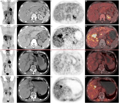
 Two cases of pathologically confirmed intrahepatic cholangiocarcinomas in 68-year-old male patients. In both cases (A-H, case 1; I-P, case 2), CT scans (B, F, case 1; J, N, case 2) revealed intrahepatic bile duct dilation and suspected nodular lesions. While F-18 FDG PET/CT (A, C, D, case 1; I, K, L, case 2) failed to demonstrate abnormal tracer uptake (SUVmax 3.5, TBR 1.3 in Case 1; SUVmax 2.6, TBR 1.2 in Case 2), F-18 FAPI-04 PET/CT (E, G, H, case 1; M, O, P, case 2 ) showed positive uptake in the nodular lesions (SUVmax 9.7, TBR 3.5 in Case 1; SUVmax 9.6, TBR 3.3 in Case 2). Image available for republishing under Creative Commons license (CC BY 4.0 DEED, Attribution 4.0 International) and courtesy of BMC Cancer.
On further analysis, F-18 FAPI-04 displayed significantly elevated SUVmax in benign liver lesions, HCCs, and ICCs, with high SUVmax correlating with lesions with hepatocyte negativity (p = 0.023), CD34 negativity (p = 0.044), and high Ki67 expression (> 30%) (p = 0.001), the researchers reported.
“This study observed a close positive correlation between F-18 FAPI-04 SUV and Ki67 expression, which is a noteworthy finding,” the group wrote.
Ultimately, beyond conventional visual analysis, semiquantitative analysis utilizing metabolic parameters such as SUV and TBR is crucial for PET/CT tracer comparison studies, the group wrote. The significance of this study is that it is among the few so far to delve into the relationship between FAPI metabolic parameters and various immunohistochemical indicators of hepatic malignancies, they noted.
“Further research is required to validate these findings and their clinical significance,” the group concluded.
The full study is available here.


