
Prostate-specific membrane antigen (PSMA) PET/CT offers superior accuracy for local staging in men with intermediate-risk or high-risk prostate cancer, according to a study published February 5 in the Journal of Nuclear Medicine.
The finding is from a head-to-head comparison of the technique with gastrin-releasing peptide receptor (GRPR) PET/CT, multiparametric MRI (mpMRI), and combined PET/CT plus MRI, noted lead author Yujia Li, MD, of Central South University in Changsha, China, and colleagues.
“PSMA PET/CT, particularly when combined with mpMRI, improves the accuracy of local staging in primary [prostate cancer] and provides independent prognostic significance beyond clinicopathologic parameters,” the group wrote.
Prostate cancer is the second most common malignancy in men worldwide and a leading cause of cancer-related mortality. Accurate local staging is crucial for guiding therapeutic strategies, including radical prostatectomy, external radiotherapy (with or without hormone therapy), focal therapy, and neoadjuvant therapy, the authors explained.
Current imaging methods, including MRI and PET/CT, may have variable accuracy in detecting key disease features, and thus the researchers aimed to determine the best approach among five standard and experimental techniques: mpMRI, PSMA-PET/CT, GRPR-PET/CT, PSMA-PET/CT plus mpMRI, and GRPR-PET/CT plus mpMRI.
The group analyzed data from 81 patients who underwent one or more of the techniques (all patients underwent mpMRI) before radical prostatectomy and compared imaging findings with whole-mount histopathology for local T stage, bilateral intraprostatic disease (BID), extraprostatic extension, and seminal vesicle invasion.
According to the results, PSMA-PET/CT showed higher overall accuracy than GRPR-PET/CT (56% vs. 36%, p = 0.011) and improved detection of BID compared with mpMRI (72% vs. 54%, p = 0.024). In a subgroup with pure acinar adenocarcinoma (AA), PSMA-PET/CT also outperformed both mpMRI and GRPR PET/CT for overall accuracy (58% vs. 39% and 34%, p = 0.029 and 0.005).
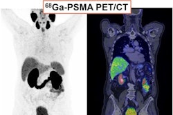
 Representative cases. (A) Patient with index lesion (AA) in right peripheral zone, scoring as Prostate Imaging-Reporting and Data System 5 on mpMRI (arrows). Two MRI readers reported discordant findings regarding lesion’s stage (1 T3a and 1 T2a). In contrast, 2 PET/CT readers correctly identified intensely PSMA-positive grade group 5 lesion, with activity extending outside gland contour, indicating EPE (miT3a). Additionally, PET/CT readers correctly identified PSMA- and GRPR-positive grade group 2 lesion (arrowheads) in contralateral site (BID on PSMA-PET/CT), which was not noted by MRI readers. (B) Patient with index lesion (ductal adenocarcinoma) in the transition zone, scoring as Prostate Imaging-Reporting and Data System 5 with bilateral involvement (BID) on mpMRI (arrows). Two MRI readers correctly identified local stage as T2c. Two PET/CT readers also accurately identified lesion on GRPR-PET/CT but failed to note BID on PSMA imaging, which showed positive nodule confined to left transition zone (miT2a). ADC = apparent diffusion coefficient; DWI = diffusion-weighted image; GG = grade group; T2W = T2-weighted. Journal of Nuclear Medicine
“This multimodal, head-to-head study demonstrates that PSMA PET/CT is a valuable tool for local staging, particularly in pure AA patients and for detecting T2c disease (BID),” the group wrote.
Ultimately, while mpMRI remains the current standard for local staging, mpMRI often underestimates disease extent, and PSMA-PET/CT is being increasingly applied for staging, the researchers noted. This study further supports its integration into clinical practice to enhance patient stratification and inform individualized treatment planning, they suggested.
“Accurate risk predication via precise staging is crucial for guiding treatment and optimizing prognosis,” the researchers wrote.
The full study is available here.


![A 53-year-old patient (patient number four) with a recurrent pituitary adenoma with extension of a cystic component of disease to the medial temporal lobe apparent on MRI (contoured in blue), and extension of disease to the left sphenoid bone and orbital apex apparent on [68Ga]Ga-DOTA-TATE (contoured in yellow).](https://img.auntminnie.com/mindful/smg/workspaces/default/uploads/2026/04/pituitary-tumor.QGsEnyB4bU.jpg?auto=format%2Ccompress&dpr=2&fit=crop&h=167&q=70&w=250)


