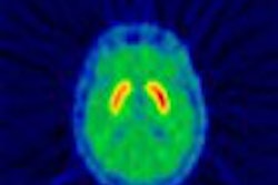
Although imaging offers a great deal of insight into the signs of dementia, it is still not precise enough for routine clinical use, according to two researchers at the 2003 Rotman Research Institute conference in Toronto.
A major difficulty is separating the brain changes associated with normal healthy aging from those that indicate the onset of pathology, said Rotman neuroscientist Cheryl Grady, Ph.D.
Fellow Rotman researcher and neurologist Dr. Sandra Black said the range of imaging techniques is expanding, and their potential applications to clinical practice are promising, "but it’s still very much a work in progress."
Most imaging studies are like the proverbial blind men who try to describe an elephant based on their sense of touch -- depending on which area they grab, they have a different impression of the animal, Grady said.
In the same way, researchers studying aging, dementia, and memory often focus on single regions of the brain, and miss how those regions are interconnected. "We need to look at the networks," Grady suggested.
Grady’s group has been working on delineating how the connections between different areas of the brain differ among young adults, healthy older adults, and people with various forms of dementia. She described two experiments with PET: One looked at the "functional connectivity" between the hippocampus and the prefrontal cortex in young and old healthy adults, while they were engaged in a memory-encoding task. The second study examined the links to the prefrontal cortex in Alzheimer’s patients during semantic and episodic memory tasks.
Grady noted that activity levels in the hippocampus during encoding of memories is correlated to the ability to recover the memories later -- a phenomenon that has been dubbed the "subsequent memory effect. But what about the larger network? We’re assuming the hippocampus is not acting alone," she said.
In the first experiment, young and healthy older adults were shown words or pictures -- such as that of a cake or a canary -- and were asked to indicate whether the subject of the picture was alive or not. There was no difference in performance, when the subjects were later asked to recognize which was which.
As Grady said she had expected, the hippocampus was correlated to performance in both groups, but there was a significant difference in what other areas were involved. The young subjects appeared to mainly use the inferior temporal regions and the anterior regions of the prefrontal cortex, while the older adults seemed to use the dorsolateral prefrontal regions and dorsal regions of the parietal area.
"It’s as if in the older adults, the hippocampus is communicating with these more organizational or executive regions, compared to the frontal regions in the young adult, and it’s this pattern which is supporting performance," she said.
The second experiment was influenced by earlier work, which has shown that Alzheimer’s patients have increased prefrontal activity and also more correlation among the prefrontal regions.
The patients and controls were also asked to perform the same living/non-living task, but in addition were studied while they were shown two words or pictures and asked to recall which they had been shown previously.
On both tasks, the healthy controls activated similar networks, Grady said, involving mainly the left hemisphere. However, the Alzheimer’s patients activated a "wide swath" of the prefrontal cortex, crossing to the right dorsolateral region, she said.
"The network in controls was pretty much limited to the left hemisphere region, but in the patients we see increased prefrontal connectivity between the left and the right," Grady said.
Patients who had a wider prefrontal network also did better on the memory tests.
In general, it appeared that the ability to use dorsolateral prefrontal regions is related to better cognitive performance, both in healthy aging and in dementia, and may represent a "general response" to change in brain function, Grady said.
"This supports the idea that there is compensatory use of alternative brain networks in older adults," she added.
While the work does show clear differences between patients and controls, imaging techniques are still only of limited use in diagnosis and prognosis in individuals. "Differential diagnosis (among different types of dementia) might be possible now, but prediction is much harder," she said.
In a separate talk at the conference, neurologist Black surveyed structural or functional imaging techniques to determine if they can detect Alzheimer’s disease, distinguish it from other dementia, diagnose the disease, predict dementia, discern changes over time, or monitor response to treatment.
Black said the range of imaging techniques is expanding, although CT is still the workhorse of structural imaging, while SPECT plays the same role in the functional area. Black said she did not find one modality more valuable than another. "As a clinician, I’ll take whatever works," she said.
However, the issue at hand is that an imaging modality that works best for research purposes -- finding differences among groups, often using automated methods -- may not necessarily have clinical applications.
"Diagnosis and prognostication require individual data," Black said. "Although we’re making progress, the answers are still not completely clear."
By Michael SmithAuntMinnie.com contributing writer
April 29, 2003
Related Reading
PET reveals hippocampal role in preserving semantic memory, April 16, 2003
Silent brain infarcts increase risk of dementia, March 27, 2003
FDG-PET spies early decline in forgetful folks, October 22, 2002
Copyright © 2003 AuntMinnie.com


