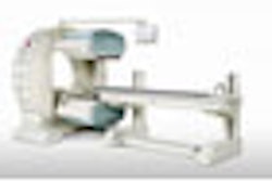
A prototype 256-slice multidetector-row CT (MDCT) scanner may be just as accurate as SPECT imaging for detecting atherosclerotic obstructions, according to data presented last week at the 2007 scientific sessions of the American Heart Association (AHA) in Orlando, FL.
Physicians with Johns Hopkins University in Baltimore suggested that MDCT perfusion images may detect obstructive arteriosclerosis with accuracy comparable to SPECT imaging. A combination of MDCT and SPECT allows clinicians to avoid the "high false-positive rate seen with SPECT imaging alone, in addition to improving prediction of territorial ischemia," they reported.
Dr. Richard George, an assistant professor of cardiology at Johns Hopkins University School of Medicine, presented the findings in a session on November 6.
"The purpose of our study was to test the feasibility of performing rest and stress 256 x 0.5-mm high-resolution MDCT perfusion imaging in patients with a history of perfusion abnormalities," George explained.
"This was a prospective clinical study that included patients with a suspected abnormal artery disease. Patients were excluded if they were medically unstable or had contraindications which prohibited use of beta-blockers, contrast agents, or adenosine," he added.
For this study, George and his team enrolled 19 patients who had abnormal SPECT perfusion studies, administering 140 µg/kg/min of adenosine for five minutes followed by rest MDCT angiography.
George noted that he and his colleagues performed the tests utilizing a prototype 256 x 0.5-mm high-resolution MDCT scanner (Aquilion 256, Toshiba America Medical Systems, Tustin, CA).
The investigative team constructed CT perfusion images of patients in the short axis with a 3-mm slice thickness, defining endocardial and epicardial borders. They divided the myocardium into sectors according to the standard 17-segment model, dividing circumferentially into endocardial and epicardial layers.
The researchers calculated the transmural perfusion ratio (TPR) by dividing the endocrinal attenuation density by the epicardial attenuation density in each sector, defining ischemia as TPR less than 0.8 in more than one sector, and comparing TPR results to the presence or absence of stenoses (≥ 50%) at CT angiography, as well as perfusion deficits on SPECT.
The group found that TPR in abnormal and normal sectors was 0.71 ± 0.005 and 1.01 ± 0.06, respectively, with the mean number of abnormal sectors for patients with no stenoses, one-vessel stenoses, and multivessel stenoses being 1.6, 2.5, and 6.3, respectively, with a statistical significance of p < 0.05.
The sensitivity and specificity of TPR for stenoses detection (≥ 50%) was 62% and 86%, respectively, compared to 62% and 71% for SPECT.
Additionally, they observed an increase in sensitivity, specificity, and positive and negative predictive values (75%, 95%, 75%, and 95%, respectively) when they defined "territorial ischemia as a SPECT perfusion abnormality plus a stenosis of greater than or equal to 50%," combining stress and rest images, the group reported.
Based on this preliminary study, the researchers are optimistic of the role of this high- resolution CT tool for detecting the presence of obstructive arteriosclerosis, but also suggest further research.
"I think in terms of future research we need to see some multicenter trials to confirm these findings," George said. In terms of the clinical implications of the study, however, he sees the use of high-resolution MDCT as a potentially valuable means for reducing radiation dose and reduction of overall contrast dose.
By Jerry Ingram
AuntMinnie.com contributing writer
November 13, 2007
Related Reading
256-slice CT brings new possibilities in head imaging, May 28, 2007
256-slice CT shows promise in one-beat, whole-heart imaging, December 7, 2006
CT pioneer test-drives 256-slice scanner, August 21, 2006
Prototype 256-slice CT scanner creates high-res images in a heartbeat, March 14, 2006
Motion-free heart images revealed with 256-slice CT, October 24, 2005
Copyright © 2007 AuntMinnie.com


