Although mammography has long been the gold standard for breast cancer screening, it does have limitations for women with dense breast tissue, with some studies reporting its sensitivity to be less than 50%.
Clinicians continue to explore other imaging techniques for this subset of women, including nuclear medicine. In fact, single-head cadmium zinc telluride (CZT) gamma cameras have shown good sensitivity in the detection of small lesions. But sensitivity still could be improved for women with large breasts or for those who have low uptake of radiotracer.
In what they believe is the first clinical study conducted using a dual-head configuration of a molecular breast imaging system, researchers at the Mayo Clinic in Rochester, MN, explored whether the use of this kind of system would increase the sensitivity of this modality (American Journal of Roentgenology, December 2008, Vol. 191:6, pp. 1805-1815).
Dr. Carrie Hruska and colleagues included 150 patients with BI-RADS category 4 or 5 lesions of less than 2 cm in the study. The team used prototype CZT detectors (GE Healthcare, Chalfont St. Giles, U.K.), which consisted of an 80 x 80 array of CZT elements with a pixel size of 2.5 x 2.5 mm, for a total detector area of 20 x 20 cm. The group also used LumaGem 3200s detectors (Gamma Medica-Ideas, Northridge, CA), which comprised a 96 x 128 array of CZT elements with a pixel size of 1.6 x 1.6 mm, giving a total detector area of 15 x 20 cm.
The molecular breast exams were read by three blinded readers and included detector 1 images only, followed by dual-head data from both detectors.
Of the 150 women included in the study, 245 lesions were evaluated. Sixty-five patients had breast density of less than 50%, 73 had breast density of more than 50%, and 12 patients had unknown breast density because mammography had not been performed or because it had been done at an outside institution. Of the patients for whom complete follow-up was available (149), 128 lesions were confirmed as cancer in 88 patients.
Sensitivity of molecular breast imaging as a function of tumor histopathology for each of three blinded readers
|
||||||||||||||||||||||||||||||||||||||||||||||||||||||||||||||||||||||||||||||||||||||||||||||||||||||||||||||||||||||||||||||||||||
| A total of 128 tumors were present in 88 patients with breast cancer. TP = true positive, FN = false negative, IDC = invasive ductal carcinoma, DCIS = ductal carcinoma in situ, and ILC = invasive lobular carcinoma. Courtesy of the American Roentgen Ray Society. | ||||||||||||||||||||||||||||||||||||||||||||||||||||||||||||||||||||||||||||||||||||||||||||||||||||||||||||||||||||||||||||||||||||
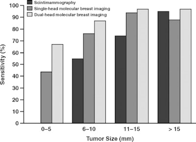
The dual-head molecular imaging studies found an average of 115 of the 128 cancers for an average overall sensitivity of 90%, while the sensitivity from single-head molecular breast imaging was 80% (102 of 128). Sensitivity of molecular breast imaging for lesions less than 10 mm was 82% for dual-head and 68% for single-head.
 |
| Dark-gray bars represent scintimammography, medium-gray represent single-head molecular breast imaging, and light-gray represent dual-head molecular breast imaging. Courtesy of the American Roentgen Ray Society. |
Hruska's team attributed the increase in sensitivity from single-head to dual-head molecular imaging to the fact that the addition of a second opposing detector decreases the maximum distance between the lesions and either detector to half the total compressed breast thickness.
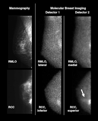
"We believe that acquiring [opposing] views from both detectors 1 and 2 provides important additional information that aids in reading the molecular breast imaging examinations and detecting very faint breast lesions," the authors wrote.
 |
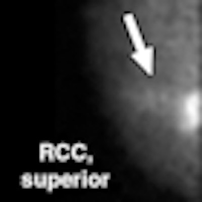
| Forty-six-year-old woman with two foci of mixed invasive ductal carcinoma with ductal carcinoma in situ in upper inner right breast: One is located at 1-o'clock position 7 cm from nipple and second is located at 2-o'clock position 3 cm from nipple. Cancers were initially identified on mammography and sonography as BI-RADS category 5 lesions. Screening mammogram and craniocaudal and mediolateral oblique views from both molecular breast imaging detectors are shown. During blinded readings, when inferior and lateral molecular breast imaging views acquired with detector 1 (middle images) were available for interpretation (corresponding to views from single-head molecular breast imaging), one focus of cancer was detected. When superior and medial molecular breast imaging views from detector 2 were available in addition to detector 1 views during separate blinded reading session, same cancer was identified but average lesion uptake score was increased from 3 to 5, indicating increase in reader confidence in identifying lesion. Also, with additional detector 2 views, second focus of cancer (arrow) was identified as blush of low-intensity uptake (uptake score of 2) visible in superior craniocaudal view only. RMLO = right mediolateral oblique, RCC = right craniocaudal. All images courtesy of the American Roentgen Ray Society. |
The team acknowledged that taking opposing views of the breast certainly can be accomplished with a single-head detector via separate acquisitions, but they stressed that acquiring the views this way doubles the imaging time, which can affect a patient's tolerance for the procedure. The researchers also acknowledged that adding a second detector head would increase the cost of the molecular imaging system, suggesting that a true cost-benefit ratio of a commercial dual-head unit compared to a single-head device still needs to be determined.
By Kate Madden Yee
AuntMinnie.com staff writer
November 28, 2008
Related Reading
Molecular breast imaging offers promise as mammography adjunct, September 15, 2008
Solid-state scintimammography matches breast MRI, July 2, 2008
Breast gamma imaging spots DCIS better than mammo, MR, August 13, 2007
Solid-state dual-head camera supercharges molecular breast imaging, December 19, 2006
Molecular breast imaging sensitive for detecting smallest tumors, January 21, 2005
Copyright © 2008 AuntMinnie.com


