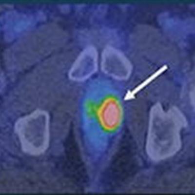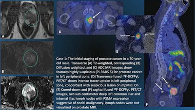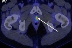
PET imaging with the radiotracer F-18 DCFPyL detects more metastatic tumors and can significantly improve treatment decisions compared with conventional imaging in patients with prostate cancer, according to research presented Wednesday at RSNA 2021 in Chicago.
Researchers at University of California, Los Angeles (UCLA) led by radiologists Dr. Soheil Kooraki and Dr. Gholam Berenji compared the performance of F-18 DCFPyL-PET/CT and multiparametric MRI (mpMRI) for initial staging of men with high-risk prostate cancer and found imaging with the tracer detected more nodal and distant metastases and change treatment in approximately 44% of patients.
"PSMA PET is more sensitive in identifying nodal and distant metastases at initial staging of men with unfavorable intermediate or high-risk prostate cancer," Kooraki said.
F-18 DCFPyL (Pylarify, Lantheus), was approved by the U.S Food and Drug Administration on May 27, 2021, for the initial staging of high-risk cancer patients for therapy or in patients whose cancer appears to have returned after radiotherapy or radical prostatectomy.
In this study, Kooraki and colleagues analyzed data from an earlier open-label, prospective phase II trial in 150 patients that explored clinical applications of F-18 DCFPyL PET/CT. They identified 41 patients from the VA Greater Los Angeles Healthcare System who had also undergone mpMRI scans.
PET/CT scans were interpreted by two nuclear medicine physicians with more than 10 years of experience. To assess changes in patient management, referring physicians were asked to complete surveys within two weeks before and four weeks after reviewing the results of the F-18 DCFPyL PET/CT scans.
Pelvic and distant lymph nodes were detected in 31.7% (13/41) and 7.3% (3/41) of individuals by F-18 DCFPyL PET/CT versus regional lymph nodes in 2.4% (1/41) on mpMRI. Skeletal metastasis was detected in 17.1% (7/41) by F-18 DCFPyL-PET/CT compared with 2.4% (1/41) by mpMRI, the researchers found.
| Disease location | mpMRI | F-18 DCFPyL-PET/CT |
| Pelvic lymph node | 2.4% (1/41) | 31.7% (13/41) |
| Distant lymph node | N/A | 7.3% (3/41) |
| Skeletal metastasis | 2.4% (1/41) | 17.1% (7/41) |
"Overall, based on the results of F-18 DCFPyl PET/CT scan, staging was altered in 44.1% of patients," Kooraki said.
 Image courtesy of Dr. Soheil Kooraki.
Clinicians at UCLA are performing targeted imaging biopsies by combining F-18 DCFPyL-PET/CT and mpMRI at the same time during initial staging to improve detection of tumors in these patients, Kooraki said. In addition to localization of intraprostatic lesions, F-18 DCFPyL- PET/CT has the advantage of depicting nodal and skeletal metastasis, he said.
"Acquiring DCFPyL PET/CT and MRI together can be beneficial for patient care in the diagnostic path of prostate cancer," Kooraki said.
Ultimately, however, long-term follow-up will be needed to determine whether this translates to better clinical outcomes, he concluded.


