ANAHEIM, CA - Although inflammation contributes substantially to the development and clinical expression of atherosclerosis, detection in humans is currently limited to indirect measurement via serum markers in the blood.
While studies have shown increased levels of serum markers of inflammation to be associated with increased risk in patients with no coronary artery disease, the sensitivity and specificity of these serum markers is uncertain. Moreover, many serum markers of inflammation are elevated in the presence of inflammatory processes not associated with atherosclerosis.
Several investigations have now indicated that MRI can detect vascular inflammation in vivo, according to a presentation at the 49th Annual Scientific Session of the American College of Cardiology this week.
Clifford R. Weiss from the National Heart, Lung, and Blood Institute (NHLBI) in Bethesda, MD, noted that MRI can detect gadolinium-DTPA contrast enhancement of the vertebral and carotid arteries in patients with suspected cerebrovascular disease -- a phenomenon believed to be associated with neovascularization of the atherosclerotic plaque. MRI has also demonstrated aortic wall thickening and gadolinium-DTPA contrast enhancement that correlates with elevation in C-reactive protein in patients with Takayasu's arteritis.
Weiss then discussed his group's latest investigation in this area. "We performed MRI studies on a 1.5-tesla cardiac scanner using a double inversion-recovery fast spin-echo sequence to assess the ability to detect vascular inflammation in vivo as defined by three potential imaging characteristics of vascular inflammation," Weiss said.
Arterial wall enhancement with gadolinium-DTPA was considered an index of intramural vasa vasorum dilation and increased capillary permeability. Increased T2 values were regarded as an index of arterial wall edema. Increased wall thickness was said to be an index of arterial wall edema and infiltration.
"Our population consisted of 27 subjects," said Weiss, "20 men and 7 women; all over 40 years of age. Ten had known coronary artery disease while 17 were healthy. Of the latter, six had elevated LDL while 11 had normal lipids and no other modifiable risk factors."
The scientists measured serum markers of inflammation, including interleukin-6, E-selectin, vascular cell adhesion molecule (VCAM-1), and intracellular adhesion molecule (ICAM-1). They also assessed C-reactive protein and obtained lipid profiles for each subject. The carotid arteries and infrarenal abdominal aorta were scanned in all subjects.
"MRI scans were categorized as abnormal if any of the MRI parameters measured -- the infrarenal aorta or the left or the right carotid artery -- was greater than one standard deviation from the population mean," Weiss explained. "We measured wall thickness, T2-weighted signal intensity, and post-contrast signal intensity. The subjects were then divided into normal and abnormal MRIs and the levels of the serum markers of inflammation in the two were compared using a two-tailed, unpaired t test."
What the NHLBI researchers found was that 15 subjects -- including the 10 with known coronary artery disease, as well as five normals -- showed one or more MRI indices of vascular inflammation in the walls of a carotid artery or the infrarenal aorta. Eight had increased T2 values or contrast enhancement without increased wall thickness; four had increased wall thickness only and three had combinations of indices.
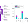
"The 15 subjects who showed indices of inflammation on MRI also had higher levels of VCAM-1, ICAM-1, and interleukin-6, compared with the remaining subjects," Weiss said. "Although the levels of C-reactive protein and E-selectin were also elevated in the abnormal MRI group, they did not reach statistical significance."
Based upon the results of their investigation, Weiss and associates concluded that MR image characteristics of vascular inflammation could stratify patients into groups of low and high markers of inflammation in healthy middle-aged and older subjects without conventional risk factors for atherosclerosis.
"Since 15 subjects had abnormal MRI markers of inflammation without physical plaque or increased wall thickness, MRI may have identified an early stage of atherosclerosis that is not available to a more morphologically based imaging technique," Weiss suggested. "Further investigation into the utility of these MRI markers is currently being performed in a larger population of subjects at risk for vascular inflammation, as well as in younger, healthy individuals," the researchers concluded.
By Sheldon M. Stern
AuntMinnie.com contributing writer
March 17, 2000
Let AuntMinnie.com know what you think about this story.
Copyright © 2000 AuntMinnie.com