Brain researchers have long sought data on the anatomical connections between parts of the human brain, relying primarily on postmortem investigation. But in vivo information on the inner workings of the brain is harder to produce.
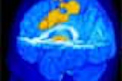
In a study they believe is the first of its kind, investigators from the U.K. explore the connections between the human thalamus and cortex using diffusion imaging data and what they call a probabilistic tractography algorithm (Nature Neuroscience, July 2003, Vol. 6:7,pp.647-781).
Diffusion tensor imaging (DTI) has been used to explore white matter of the brain in living human subjects, but not necessarily to map the connections between the brain’s gray matter like the thalamus and cortex. The thalamus in particular is key to understanding brain function, as it sifts the majority of incoming information to the cortex.
But the structure of the thalamus makes its function difficult to image, even with technology such as MRI. Diffusion imaging differentiates the diffusion properties of water. So in tissue with "a high degree of directional organization," like that of the brain, it is possible to map the diffusion of water protons as they disperse in different directions, according to the authors, who hailed from the University of Oxford in Oxford and University College in London.
Using echo-planar imaging with a 1.5-tesla Signa Horizon scanner (GE Medical Systems, Waukesha, WI), the team collected data from eight subjects (six men and two women, ages 26 to 33). The majority of the results reported in the article come from data gathered from a 33-year-old male subject.
Conventional "streamlining" tracts, or the algorithms that are traditionally used with DTI, work best when the fiber direction of brain tissue is certain. The group’s probabilistic tractography algorithm "infers anatomical connectivity that progresses fully into gray matter," according to the article, providing the additional advantages of predicting the relative probability of fiber directions and resisting noise in the image.
Using this algorithm, the researchers traced the pathways from the thalamus to the cortex, finding that the connections between the two were comparable to those found in studies of non-human primates. This similarity was not limited to the distribution of connections to different cortical sites, but also was found in the pathways that are taken between the thalamus and the cortex.
The researchers said that the methods they used in this study could have clinical applications for tracking developmental and acquired brain disorders by testing for variations in frontothalmic circuitry in schizophrenia, or exploring quantitative differences in brain connectivity and learning abilities. It could also help clinicians more accurately target brain structures in the treatment of movement disorders with functional neurosurgery, according to the article.
By Kate Madden YeeAuntMinnie.com contributing writer
July 16, 2003
Related Reading
Brain MR scans validate individualized pain ratings, July 8, 2003
Alzheimer's brains show features of immaturity, May 8, 2003
NIH licenses diffusion tensor MRI technology, August 5, 2002
Copyright © 2003 AuntMinnie.com


