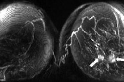
VIENNA - Radiography and MRI turn in similar sensitivities for assessing joint erosion in rheumatoid arthritis (RA), although MRI may ultimately prove to be the superior modality, according to a presentation Thursday at the European Congress of Rheumatology.
The researchers suggested that MRI may be useful in evaluating the treatment effects of the newer biologic agents, perhaps enabling clinicians to assess changes in joint damage earlier.
"As MRI becomes cheaper or more accessible, then you can use it to evaluate patients during biologic therapies. You can see which patients respond to treatment, and you can change the treatment according to your MRI assessment," explained Espen Haavardsholm, a doctoral candidate from the department of rheumatology at Diakonhjemmet Hospital in Oslo, Norway.
For this preliminary investigation, Haavardsholm and colleagues enrolled 27 patients diagnosed with clinically active RA. Clinicians evaluated core measures of disease activity using MRI and x-ray once patients began treatment with one of three anti-TNF agents (etanercept, infliximab, or adalimumab).
They assessed patients at three, six, and 12 months during the course of treatment. MRI evaluations were done on a 1.5-tesla unit. They scanned the dominant wrist following the core set recommended by the Outcome Measures in Arthritis Clinical Trials (OMERACT), Haavardsholm reported. Anterioposterior radiographs using high-resolution digital techniques were taken of both hands and wrists.
MR images were analyzed according to the RA-MRI scoring system (RAMRIS, 0-150). For x-ray assessment, the researchers used the van der Heijde-modified Sharp score (range 0-160).
They examined changes in annual progression rates of joint erosion using a paired samples t-test. Finally, researchers assessed the sensitivity to change of the two scoring methods by calculating the standardized response mean (SRM).
The investigators found similar sensitivities in MRI and conventional radiograph scoring systems with an SRM ratio of almost 0.8. SRM for x-ray was -0.75 and -0.78 for MRI. They also found concordance between the two methods in approximately 40% of the cases.
While the results are promising, Haavardsholm stressed the small size of the patient cohort. He said that larger studies are under way that will test the impact of biologic therapies, as well as the best means of evaluation.
By Jerry Ingram
AuntMinnie.com contributing writer
June 10, 2005
Related Reading
MRI and CT: Complementary techniques in assessing rheumatoid arthritis. March 9, 2005
The twists and turns of hand and wrist x-ray positioning, October 15, 2002
Copyright © 2005 AuntMinnie.com


