Parallel imaging in breast MRI promises to enhance the efficiency of data postprocessing and improve exam times. The process takes advantage of phased-array coils operated by multiple receivers, reducing the need for magnetic gradient switching and resulting in faster scanning without the loss of image resolution.
For breast MRI exams this procedure allows the imaging of both breasts simultaneously. This translates to reduced motion artifact and improved image quality, according to a presentation at the 2006 American Roentgen Ray Society (ARRS) meeting in Vancouver, British Columbia.
A group from the department of radiology at Northwestern University's Feinberg School of Medicine in Chicago utilized a pair of reconstruction algorithms for its parallel breast MRI studies, which were compared to nonparallel imaging techniques.
The team used generalized autocalibrating partially parallel acquisitions (GRAPPA), an enhanced version of simultaneous acquisition of spatial harmonics (SMASH), which is a reconstruction algorithm that operates on partial k-spaces, one from each coil, prior to image generation by fast Fourier transformation (FFT).
The group also employed modified sensitivity encoding (MSENSE), an enhanced version of the SENSE reconstruction algorithm that operates on partial images from each coil that have been generated by FFT. Both algorithms are part of Siemens Medical Solutions (Malvern, PA) integrated parallel acquisition techniques (iPAT) software for MRI reconstructions that the scientists used to conduct their study.
"Simply put, breast imaging with parallel acquisition uses multichannel coils and knowledge of each coil's sensitivity volume to collect two or more channels of data simultaneously," said Dr. Lora Barke, who presented the results of the group's research.
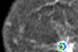
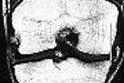
The researchers performed contrast-enhanced bilateral phantom, volunteer, and clinical breast MRI studies with and without parallel imaging on Siemens Sonata, Symphony Quantum, and Avanto 1.5-tesla MRI systems. They utilized a 3D Fourier transformation (3DFT) gradient-echo technique that employs computer processing to combine individual slice acquisitions together to produce an image that represents length, width, and height. A minimum time to rotate (TR) of 3.8 to 4.6 msec and time to echo (TE) of 1.6 to 1.8 msec was used to obtain fat-suppressed images with nearly isotropic voxels, according to Barke.
"We used a four-channel breast coil; the sensitivity profile of each coil was measured, and the datasets for both breasts were collected simultaneously," she said.
Temporal resolution was held below 120 seconds for both parallel and nonparallel techniques. The resulting images from both techniques were compared in terms of acquisition time, signal-to-noise ratios (SNR), image artifacts, and overall image quality, Barke said.
Parallel imaging with either GRAPPA or MSENSE provided an acceleration factor of 2 without a loss of spatial resolution. As the acceleration increased, the SNR decreased, according to Barke.
"With all spatial parameters kept constant, parallel imaging incurred a loss of SNR of approximately 35% to 45% without significant increase in artifacts in the axial or coronal plane," she said.
However, the group did discover that parallel imaging with primary acquisitions in the sagittal plane caused significant artifacts. Barke noted that with isotropic or nearly isotropic 3D acquisitions in the axial or coronal plane that postacquisition reconstruction could be performed in the sagittal or oblique planes, along with maximum intensity projections (MIPs), without artifacts and without significant loss of spatial resolution.
The exam time was nearly cut in half through the use of parallel imaging. For the nonparallel studies, the exam time was approximately 21 minutes. With parallel imaging at an acceleration factor of 2, it dropped to almost 13 minutes, Barke said.
The researchers found that applying parallel imaging to breast MRI with currently available commercial software improved temporal resolution without loss of spatial resolution and without significant loss of image quality or a significant increase in image artifacts.
"There is a restriction in the primary acquisition to the axial or coronal planes, but reconstruction can be performed in any projection," Barke said.
By Jonathan S. Batchelor
AuntMinnie.com staff writer
July 13, 2006
Related Reading
Breast MR falls short for microcalcifications, but CAD helps in malignancies, July 3, 2006
MR spectroscopy aids breast cancer diagnosis, June 8, 2006
Technology, guidelines cement role for breast MR in cancer screening, May 26, 2006
MR mammography: Screening tool or diagnostic adjunct? February 9, 2006
Larger lesions on breast MRI equal greater chance of cancer, February 8, 2006
Copyright © 2006 AuntMinnie.com