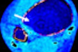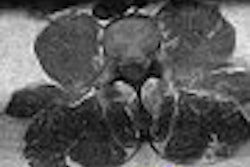
A parallel imaging technique combined with 3-tesla MR can reduce motion artifacts, cut down on scan time, and allow for a flexible scanning protocol for ankle assessment, according to a paper in the July issue of the American Journal of Roentgenology.
Dr. Jan Stefan Bauer, Dr. Suchandrima Banerjee, and colleagues from the University of California, San Francisco (UCSF) and the UC Berkeley/UCSF Joint Graduate Group in Bioengineering in Berkeley, CA, tested out the generalized autocalibrating partially parallel acquisition (GRAPPA) technique in cadaver specimens and healthy human volunteers. They compared imaging results for GRAPPA at 3-tesla MR and in small field-of-view (FOV) 1.5-tesla MR.
MR scans were obtained in three fresh, human cadaver specimens with abnormalities of the ligaments, tendons, and cartilage. The three volunteers had no ankle abnormalities. Scanning was done on the 1.5- and 3-tesla units (Signa, GE Healthcare, Chalfont St. Giles, U.K.) with a protocol that consisted of axial and sagittal T1-weighted, axial fat-saturated T2-weighted, coronal fat-saturated intermediate-weighted fast spin-echo (FSE) imaging. A fat-saturated spoiled gradient-recalled (SPGR) sequence was used for dedicated cartilage imaging. The scan time was approximately six minutes for each sequence at both magnet strengths.
"Partial parallel imaging was used with a reduction factor (R) of 2 and 16 calibration lines," the authors explained. "Thus, the parallel imaging datasets consisted of 144 phase encodes compared with 256 in a conventional acquisition.... For the partially parallel imaging datasets, images were reconstructed using a GRAPPA-based algorithm, whereas full imaging datasets were reconstructed using a standard Fourier construction" (AJR, July 2007, Vol. 189:1, pp. 240-245).
The signal-to-noise ratio (SNR) was measured for every sequence, and the contrast-to-noise (CNR) ratio was calculated for SPGR sequences. For parallel imaging, SNR and CNR were based on twice-repeated acquisitions. All images were assessed by two radiologists for quality (sharpness of edges, blurring and noise, delineation of ligamentous structures, tissue contrast) using a four-point confidence scale, with 4 representing an optimal image.
 |
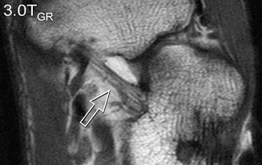 |
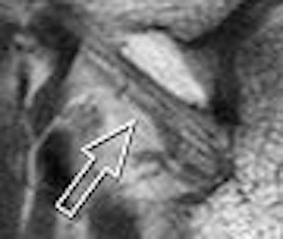
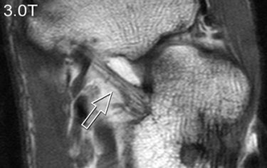
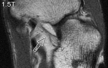
| A 31-year-old healthy male volunteer. Axial T1-weighted fast spin-echo images of foot show superior delineation of spring ligament (arrow) at 3 T (top and middle) as opposed to 1.5 T (below); 3.0TGR = 3.0T with GRAPPA algorithm. No significant difference was found between (top) and parallel (middle) acquisitions at 3 T; visualization of this ligament was rated very good at 3 T and as moderate at 1.5 T (below) by both radiologists. |
 |
| Bauer JS, Banerjee S, Henning TD, Krug R, Majumda S, and Link TM, "Fast High-Spatial-Resolution MRI of the Ankle with Parallel Imaging Using GRAPPA at 3 T" (AJR 2007; 189:240-245). |
According to the results, GRAPPA MR resulted in a 44% reduction in scan time compared with conventional imaging. SNRs and CNRs doubled on the SPGR sequence at 3 tesla. These 3-tesla images also demonstrated comparable edge sharpness.
Image quality was rated highest on 3-tesla images. The axial T1-weighted sequence was given an average score of 3.4 at 3 tesla for parallel and normal acquisitions versus a score of 2.8 for 1.5 tesla. Visualization of ligaments and tendon abnormalities were also rated highly on parallel 3-tesla MR.
The higher field strength plus GRAPPA in the ankle produced "excellent" diagnostic-quality images and a faster scan time, the authors concluded. With regard to protocol design, GRAPPA can be used with 3D sequences to improve volume coverage or with 2D FSE sequences to reduce T2 blurring, they explained.
The authors also noted that, in this application, GRAPPA has advantages over other parallel imaging techniques. Small FOV imaging suffers from aliasing while SENSE parallel imaging has problems with image reconstruction.
They acknowledged that this was a preliminary study and that a large-scale clinical study would be required. In addition, optimized receiver arrays will improve parallel imaging results.
By Shalmali Pal
AuntMinnie.com staff writer
June 29, 2007
Related Reading
MRI keeps pace with rapidly evolving musculoskeletal systems of young athletes, May 20, 2007
Studies uphold MRI's role in meniscal tears, propose new sequences, May 16, 2007
GRAPPA MRA curtails gadolinium dose in peripheral arterial occlusive disease, March 21, 2007
Copyright © 2007 AuntMinnie.com


