CT is the gold standard for lung imaging, particularly when a chest x-ray lacks the detail needed to make a diagnosis. But CT's far higher radiation dose -- equivalent to 200 chest radiographs -- has some pediatric radiologists looking for an alternative modality, especially for children with chronic lung conditions who may require many CT scans.
Radiologists at Turku University Hospital in Turku, Finland, are using MRI in many cases of problematic lung infections where a CT exam might have been performed following a routine chest radiograph. A newly published article in Pediatric Radiology (November 2008, Vol. 38:11, pp.1,225-1,231) discusses how they did it.
The study analyzed MRI exams performed between April 2005 and February 2007 in 24 pediatric patients with suspected lung infections. The research team was led by Dr. Ville Peltola, a pediatric specialist in the department of pediatrics at the university.
Indications for an MRI examination include suspicion of a complicated or chronic lung infection based on clinical findings and/or the results of a chest radiograph, or the existence of a chronic condition associated with lung complications, according to Peltola.
Approximately 10% of the 100-120 children admitted annually to Turku University Hospital for treatment of pneumonia have an MRI performed, he said. Patients with cystic fibrosis, empyema, and recurrent pneumonia or chronic lung disease, with or without documented primary immunodeficiency, are often studied with MRI, Peltola said.
The five boys and 19 girls who were the subject of the study ranged in age from one month to 15.9 years, with the majority of patients being of an age where they would not require sedation (under two months of age or older than five years). Eleven of the patients had chronic conditions predisposing them to lung infections, including asthma, cystic fibrosis, juvenile rheumatoid arthritis, cilia dysfunction, colonic aganglionosis, hypogammaglobulinaemia, and middle lobe syndrome.
The protocol at Turku University Hospital is to perform a digital chest radiograph for all children with suspected lung infections. An MRI is ordered in cases where pneumonic infiltrates, adenopathy, or complications cannot be detected in lung areas obscured by the heart, mediastinum and/or diaphragm, or when it is not possible to differentiate between sterile parapneumonic effusion and pleural empyema.
For the study, a 1.5-tesla MR scanner (Magnetom Avanto, Siemens Healthcare, Erlangen, Germany) was utilized, with a protocol consisting of coronal T2-weighted, axial T2-weighted, and coronal short-tau inversion recovery (STIR) images of the lungs. For free-breathing children, T1-weighted fat-suppressed axial and coronal images were also obtained after contrast injection. All MR images were obtained with a slice thickness of 5-6 mm.
Dr. Erkki Svedström, a pediatric radiologist and a co-author of the article, interpreted all of the studies. Images were compared to detect parenchymal, pleural, and lymph node enhancement. Contrast-enhanced fusion images were produced as required.
Special attention was also paid to identify the microbiological etiology of the patients. Sputum samples were collected of all children, and bronchoalveolar lavage, pleural fluid, and nasopharyngeal aspirate specimens were also collected, depending on the clinical presentation. This was done to better understand the MRI findings in infections caused by different microbes, Peltola explained.
The predominant MRI findings were alveolar or interstitial parenchymal changes, pleural thickening and fluid, and lymph node enlargement, which are not often detected by chest x-ray. Pulmonary abnormalities were well characterized by MRI in uncomplicated and complicated pneumonia in previously healthy children, and in subacute or chronic lung infections in patients with chronic conditions.
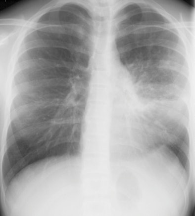 |
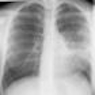
| A 13-year-old girl with pneumonia caused by Mycoplasma pneumoniae. Chest x-ray (above) and a contrast-enhanced 1.5-tesla coronal image (below). The MR image shows patchy parenchymal changes in both lungs. Images courtesy of the Department of Radiology, Turku University Hospital. |
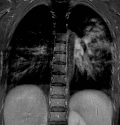 |
Contrast enhancement was detected in active infectious process. It was not detected in noninfectious lung process, such as a pleural fluid collection secondary to peritonitis. In nine of 10 patients with uncomplicated community-acquired pneumonia, pleural fluid was detected, and the pleura enhanced with contrast medium.
MRI was useful when clinicians suspected complicated pneumonia, such as empyema or lung abscess. In a related study published last year at Turku University Hospital (Acta Paediatrica, November 2007, Vol. 96:11, pp. 1,686-1,692), empyema was not detected by chest x-ray in one-third of the 37 pediatric patients studied.
Faster diagnosis and treatment of empyema may shorten the length of hospitalization and reduce the cost of treatment, as well as enable more timely surgical intervention when it is required. As yet, no length-of-stay or cost-benefit analyses have been conducted, Peltola noted.
Peltola and colleagues recommend that studies be conducted to evaluate the power of contrast enhancement to discriminate between active infection and other lung processes, as well as detection of complications, microbe-specific changes, and pathogenic processes of pneumonia.
"MRI may give new insight into the pathophysiology of pneumonia," Peltola said. "Our experience of MRI of infections and chronic lung changes in cystic fibrosis and other chronic lung conditions is limited but encouraging. Larger studies comparing MRI and CT for these indications are urgently needed."
By Cynthia Keen
AuntMinnie.com staff writer
October 16, 2008
Related Reading
CAD offers potential for diagnosing pediatric pneumonia, July 23, 2008
Grown-up decisions: When is MDCT the best choice for children? September 30, 2005
Copyright © 2008 AuntMinnie.com


