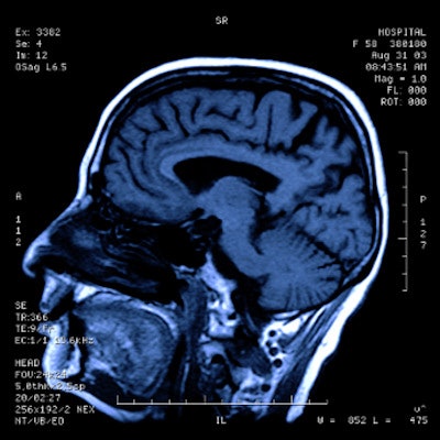
Performing advanced MRI scans on people with mild traumatic brain injury (mTBI) within 72 hours could translate to better patient outcomes, according to a study published March 18 in JAMA Network Open.
A team of researchers led by Dr. Sophie Richter of the University of Cambridge in the U.K. found that early MRI scans can identify white-matter injury associated with symptoms after mTBI and, if tracked early, could help clinicians determine whether patients are at risk of poor outcome -- and require early intervention.
"Imaging may thus document the evolution of pathology, thereby highlighting windows for therapy," the group noted. "In addition, imaging can provide prognostic information to help select patients for clinical follow-up or interventional trials."
Traumatic brain injury affects millions of people each year, and although most injuries (70% to 90%) are classified as mild, this may not be the most accurate way to describe the condition, the investigators wrote.
"This term [mild] ... is clearly a misnomer, because 30% to 50% of those patients experience symptoms that persist beyond six months and disrupt relationships and employment," they wrote.
MRI offers a way to better assess patients with mTBI at time of injury and therefore identify those who may need more intervention, according to the authors. In their study, they sought to explore three questions:
- Are there particular neuroanatomical substrates of mTBI?
- How may these substrates change over time?
- When is the best time to image with MRI in order to estimate patient outcomes?
The study included data from 81 patients who presented at the hospital within 24 hours of an mTBI (measured by Glasgow coma scores of 13 to 15) who underwent a first MRI scan within 72 hours of injury and a second scan two to three weeks later. Most of the injuries were caused by falls from a height (26%) or ground-level falls (23%), and most study participants had a Glasgow coma score of 15 (79%). The researchers also included 104 control participants for comparison.
MRI sequences included advanced data from volumetric T1-weighted, volumetric fluid-attenuated inversion recovery (FLAIR), T2-weighted, susceptibility-weighted imaging, and diffusion tensor imaging (DTI), the group wrote.
The most prominent finding was reduction in white-matter volume between the first MRI scan and the second -- which the researchers posited could be due to resolution of swelling or to later loss of white matter. On analysis, the group noted that the decreased white-matter volume suggested pathology.
"Compared with controls, patients had similar [white-matter] volumes at [the first MRI] but reduced volumes at [the second]," the team noted. "This finding suggests that the reduction of white-matter volume at [the second MRI] ... did not represent resolution of edema but rather new, persistent, and potentially progressive pathology."
| Changes in white-matter volume on MRI in patients with mild TBI | ||||
| Area of interest | First MRI | Second MRI | Absolute difference | p-value |
| Cerebral white-matter volume | 231.5 cm3 | 229.8 cm3 | -3.77 | 0.001 |
Richter's team also discovered that the "association between recovery and imaging findings was significantly closer" on the first MRI scan than at the second, with an area under the receiver operating curve measure of 0.87 compared with 0.75. The early MRI scan also showed higher positive and negative predictive value.
"Our findings demonstrate that MRI appearances are dynamic and that images obtained closer to the time of injury are more closely associated with outcome," the group wrote.
The study highlights the promise of MRI for assessing people with mild TBI, wrote a team led by Dr. Elie Massaad of Massachusetts General Hospital in Boston.
"Advanced MRI methods have shown promise for improved prognostication in mild TBI," Massaad and colleagues wrote. "[This study] provides further evidence that there is an association between structural changes detected on DTI ... and symptom severity and persistence."


