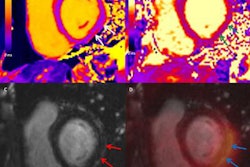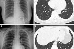
Myocarditis after COVID-19 mRNA vaccination is rare, but it does occur, and cardiac MRI helps clinicians identify it, according to a study published October 27 in the American Journal of Roentgenology.
The findings are important to consider, especially for children, since earlier this year the U.S. Food and Drug Administration (FDA) gave emergency use approval to the Pfizer vaccine for adolescents between 12 and 15, wrote a team led by Dr. Lydia Chelala of University of Chicago Medicine.
"[In] the United States occurrences of myopericarditis in adolescents and young adults after COVID-19 mRNA vaccination have ... been reported by the Vaccine Adverse Event System," the group noted. "This possible association warrants further investigation, particularly given the [FDA's emergency approval in May of the Pfizer vaccine for youth]."
Developing myocarditis after COVID-19 vaccination is uncommon, but its incidence may be underestimated because diagnosis depends on symptoms, the authors wrote. And underestimating myocarditis, particularly in young people, can be dangerous.
"A study in young athletes with confirmed COVID-19 showed that a substantial proportion of individuals had subclinical myocarditis on screening cardiac MRI," they noted. "Subclinical myocarditis may also be possible after COVID-19 vaccination ... To better understand a possible association, the CDC has encouraged further investigation of myocarditis following COVID-19 mRNA vaccination."
Chelala and colleagues identified 52 people who underwent cardiac MRI for suspected myocarditis between May and June 2021. Of these, five had the exam after having recently received an mRNA-based COVID-19 vaccine; they had no previous history of the illness. All five were boys between the ages of 16 and 19 who presented at the hospital within four days of receiving their second vaccine dose.
Troponin levels were high in all of the patients, which prompted the cardiac MRI exam. The investigators ruled out other possible causes of myocarditis using medical history information, physical exam, a myocarditis viral panel, and a toxicology screen.
Cardiac MRI exam findings indicated myocarditis in each of the five boys according to Lake Louise criteria (regional or global signal intensity increase on T2 images; increased global myocardial early gadolinium enhancement ratio; and at least one focal, nonischemic lesion at inversion-recovery late gadolinium enhancement imaging).
All the boys recovered and were discharged in stable condition after a mean hospital stay of 4.8 days. Two of the five underwent further cardiac MRI exams within a follow-up period (mean length, 95 days) that indicated continuing, though decreased late gadolinium enhancement. Three of the five experienced mild chest pain after being discharged from the hospital that eventually resolved.
As clinicians continue to better understand the effects of COVID-19 disease -- and vaccinations against it -- it's necessary to keep the possibility of myocarditis in mind, according to Chelala and colleagues.
"[Radiologists] should be aware of the possible association and recognize the role of cardiac MRI in the assessment of patients with suspected myocarditis after COVID-19 vaccination," they concluded.


