An MRI neuroimaging-based machine-learning model shows promise for helping clinicians diagnose attention deficit hyperactivity disorder (ADHD), according to research to be presented at the upcoming RSNA meeting.
The findings suggest that there is a less subjective way of identifying the condition, which could lead to better treatment planning and tracking, noted a team led by Huang Lin, PhD, of the Yale School of Medicine in New Haven, CT.
"There's a need for a more objective methodology for a more efficient and reliable diagnosis," Lin said in a statement released by the RSNA on November 23. "ADHD symptoms are often undiagnosed or misdiagnosed because the evaluation is subjective."
In the U.S., ADHD affects 6 million children between the ages of 3 and 17, the investigators noted. The condition makes it difficult for children to pay attention and control impulsive behavior. Diagnosis is a subjective process that relies on the child's physician or caregiver to identify ADHD symptoms.
Lin's group sought to explore whether brain imaging could improve the diagnosis of ADHD. They investigated whether MRI in particular would show any microstructural, morphological, and functional connectivity correlates of the disorder.
The investigators' research included 1,798 children with ADHD and 6,007 without it. They used data from the Adolescent Brain Cognitive Development (ABCD) study -- which consists of information from almost 12,000 participants between the ages of 9 and 10 -- adjusting it for age and sex and tracking neuroimaging metrics such as brain volume, surface area, white matter integrity, and functional connectivity. They also developed, tested, and validated a variety of machine-learning algorithms for predicting ADHD.
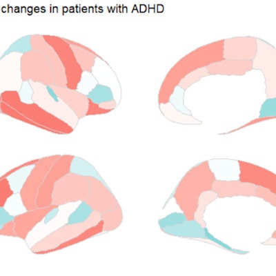
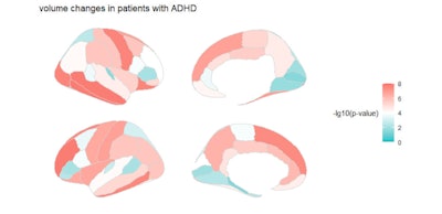
The study showed that adolescents with ADHD had abnormal connectivity in brain networks involving memory and auditory processing, a thinner brain cortex, and white matter microstructural changes in the frontal lobe.
 Volume changes in patients with ADHD. Children with ADHD tend to have lower cortical volume, especially in the temporal and frontal lobes. Image and caption courtesy of the RSNA.
"Abnormal functional connectivity, involving networks related to memory processing, tonic alertness, and the auditory process, thinning of brain cortex, and white matter tract neural fiber loss and microarchitectural disintegrity are markers of ... ADHD," the team wrote.
Of the machine-learning models, the combination of elastic net logistic regression with hierarchical clustering feature selection demonstrated the highest performance, with an area under the receiver operating curve of 0.6085 for ADHD prediction, the group reported.
The findings further support ADHD's neurological basis, according to Lin.
"Our study underscores that ADHD is a neurological disorder with neurostructural and functional manifestations in the brain, not just a purely externalized behavior syndrome," she said.


