Researchers from Children's Hospital of Philadelphia are highlighting a genitourinary 3D modeling and printing process using post-contrast MRI that they believe can help with pediatric urologic surgical planning.
A team led by Elizabeth Silvestro described its findings in research published January 11 in Urology. The group used the process to model genitourinary structures, including solid organs and body cavities, in two children.
"This technique could be extended to any time-course imaging sequence for 3D printing," the group wrote. "This application could have broad applications across [multiple disciplines] and offers new ways to visualize the imaging data to inform surgical planning."
Previous studies have described the effect of applying 3D modeling and additive manufacturing to images taken from various radiology disciplines, as well as how such modeling can help plan treatment. But Silvestro's group noted that 3D modeling and printing for urological applications hasn't been explored as much as it could be.
The group sought to test a process they developed that converts multiphase contrast-enhanced MR images into merged 3D renderings and printed models. They used MR images from a 13-month-old girl undergoing complex genitourinary anatomy reconstruction and in a 32-month-old boy with multiple Wilms tumors.
For this process, the team took multiphase gadolinium-enhanced free-breathing 3D T1-weighted sequences for MR imaging, gave them to a radiologist and an engineer to select relevant images for inclusion into the model, used a base scan from precontrast images to align anatomy, and selected the best image of each region of interest. The images were then loaded into a segmentation program, and the regions of interest were modeled using threshold and manual contouring before being exported and overlaid on the base scan and aligned, as well as edited.
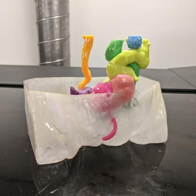
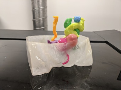 A 3D-printed model shows the genitourinary system of a 13-month-old girl. This model consists of hemibladders (pink), duplicated vaginas (purple), uterine horns (light purple), ureters (orange and green), ovaries (blue), urethra (pink), colon (yellow), and a body extent (clear). The sides of the body are flat due to the imaging field of view. Images courtesy of Urology.
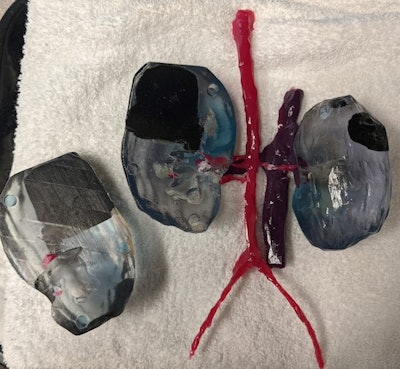 A 3D printed model depicts kidneys from a 32-month-old boy with multiple Wilms tumors. The model includes tumor (black), kidney (clear), collecting system (hollowed cavity), arteries (pink), and veins (purple). The model depicts how the tumors encroach on the kidney and collecting systems and allow for the kidney to be opened.
The 13-month-old girl had previously undergone surgical creation of a colostomy and vesicostomy. Before undergoing her MRI, during an exam under anesthesia, catheters were placed into her. The printed model the investigators created for her consisted of hemibladders, duplicated vaginas, uterine horns, ureters, ovaries, urethra, and colon.
For the 32-month-old boy, initial imaging showed tumors in both kidneys, as well as metastatic lung lesions. He started chemotherapy for presumed Wilms tumors. In his case, the printed model included the tumors, kidneys, a collecting system symbolized by a hollowed cavity, arteries, and veins; it showed how the tumors were encroaching on the kidney.
Silvestro's group consulted with urologists and two general surgeons, who reported that the models helped them better understand connections between anatomical structures and better plan surgical interventions.
"[The model] is a tangible depiction of the anatomy to aid presurgical counseling of the family," the authors wrote. "Urologists [can] use the models to explain the planned reconstruction to each patient and their family."


