Using computer-assisted detection (CAD) software to assess enhancing breast tissue as a percentage of the entire breast volume on contrast MR imaging shows promise for improving breast cancer diagnosis, researchers have found.
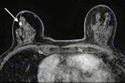
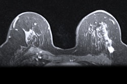
In a pilot study, a team led by Barbara Bennani-Baiti, MD, of the Medical University of Vienna in Austria, found that in 39 patients who underwent two contrast-enhanced breast MRI exams, CAD software yielded areas under the receiver operating curve (AUC) as high as 0.84. The results could help streamline workflow in busy radiology departments and perhaps even make breast MRI more accessible, according to the researchers, whose findings were published March 12 in the Journal of Magnetic Resonance Imaging.
"[A tool that] would greatly alleviate the workload and free the reporting radiologists to review the more complicated cases … would … enable wide-scale application of breast MRI exams and make [MRI] accessible in areas where dedicated breast radiologists are scarce, benefiting areas that are more rural and low-income communities," it wrote.
AI-based tools to ease radiology workflow show promise, but "often suffer from the lack of transparency concerning the methodology and features that lead to an alleged diagnostic efficiency," Bennani-Baiti and colleagues explained. The team sought to explore whether a simple CAD-based tool that uses existing software could help distinguish malignant from benign lesions on breast MRI.
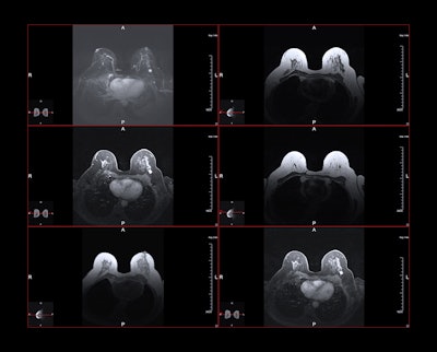
The investigators conducted a study that included 39 patients who underwent two subsequent breast MRI exams for suspicious results on conventional imaging with 0.1 mmol/kg each gadobenic acid (MultiHance, Bracco) and gadoteric acid (Dotarem, Guerbet). Two readers blinded to the cases' histopathological outcomes evaluated unenhanced and early post-contrast images using computer-assisted software (Brevis, Siemens Healthineers). Bennani-Baiti and colleagues calculated the software's diagnostic performance for percentage of ipsilateral voxel volume enhancement and for percentage of contralateral enhancing voxel volume and compared these results to histopathological results.
The group found that both approaches resulted in promising area under the receiver operating curve (AUC) values for detecting breast cancer:
| Performance of CAD software for classifying breast tissue volume and parenchymal enhancement compared with histopathological results (AUC) | |
|---|---|
| Ipsilateral enhancing voxel volume vs. histopathological outcome | |
| Gadobenic acid | |
| Reader 1 | 0.71 |
| Reader 2 | 0.69 |
| Gadoteric acid | |
| Reader 1 | 0.78 |
| Reader 2 | 0.77 |
| Background parenchymal enhancement vs. histolopathological outcome | |
| Gadobenic acid | |
| Reader 1 | 0.79 |
| Reader 2 | 0.84 |
| Gadoteric acid | |
| Reader 1 | 0.69 |
| Reader 2 | 0.66 |
The simplicity of this approach has much to recommend it, according to the authors.
"This method is easily applicable for many institutions and does not require costly specialized software packages," they concluded. "Yet it might facilitate breast MRI reading and application in less specialized centers and make breast MRI more accessible to more women as the need for breast MRI exams keeps increasing."
The complete study can be found here.