Light therapy appears to promote healing in people who have suffered significant brain injuries, researchers have found.
Patients who underwent low-level light therapy showed a greater change in resting-state connectivity in seven brain regions during the acute-to-subacute recovery phase from brain injury compared to control participants, reported a team led by Suk-tak Chan, PhD, of Massachusetts General Hospital (MGH). These results were made plain on functional MR imaging and were published May 28 in Radiology.
"There was increased connectivity in those receiving light treatment, primarily within the first two weeks," study co-author Nathaniel Mercaldo, PhD, said in a statement released by the RSNA.
The healing effect of light on the brain has been studied for years, the researchers noted. They conducted a study in which 38 patients who had suffered moderate traumatic brain injury -- serious enough to alter cognition and/or be visible on a brain scan. Of these, 17 underwent low-level light therapy within 72 hours of the injury via a helmet that emitted near-infrared light -- 810-nanometer-wavelength -- and 21 did not (thus serving as a control group). The investigators focused on the brain's resting-state functional connectivity, or the communication between brain regions that occurs when a person is at rest. They then compared MRI results from three time periods: the acute phase (within one week after injury); the subacute phase (two to three weeks postinjury); and the late-subacute phase (3 months after injury).
Brain connectivity increased for the light therapy-treated patients during the acute to subacute phases, although there was no evidence of a difference in clinical outcomes between the treated and control participants -- which suggests that additional studies with larger cohorts of patients are needed.
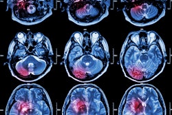
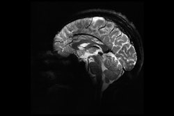
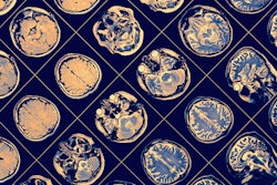
 Functional MRI brain maps of resting-state functional connectivity in representative age- and sex-matched participants. (A, B) Axial (top) and coronal (bottom) views show whole-brain connectivity, with the seed at the left (L) superior frontal region, in a 36-year-old female participant in the low-level light therapy (LLLT) treatment group (A) and a 38-year-old female participant in the sham treatment group (B) during the acute, subacute, and late-subacute phases (columns, from left to right, in both A and B) of traumatic brain injury recovery. (C) Axial (top left), coronal (bottom left), and sagittal (right) views in a 38-year-old female control participant are shown for comparison; the solid green circle in the sagittal view indicates the location of the left superior frontal seed region. The color bar indicates that brain regions with warm colors (red, orange, yellow) show resting-state fluctuations that have significant positive correlation (r of 0 to 1) with those of the left superior frontal region, and brain regions with cold colors (blue) show resting-state fluctuations that have significant negative correlation (r of −1 to 0) with those of the left superior frontal region. Brain regions that have functional connectivity with the left superior frontal seed in the LLLT-treated participant (arrowheads in A) but not in the sham-treated participant (arrowheads in B) are shown. The arrow in A additionally shows brain regions with positive correlation with the seed in the LLLT-treated participant, but negative correlation with the seed in the sham-treated participant (arrow in B). All connections shown here achieved a false discovery rate–adjusted p < 0.005. Images and caption courtesy of the RSNA.
"There is still a lot of work to be done to understand the exact physiological mechanism behind these effects," Chan said.
In any case, the study findings could translate to other ways to use light for healing in the brain, according to senior author Rajiv Gupta, MD, PhD, also of MGH.
"There are lots of disorders of connectivity, mostly in psychiatry, where this intervention may have a role," Gupta said in the statement. “[Post-traumatic stress disorder], depression, autism: these are all promising areas for light therapy."
The complete study can be found here.