Men should have prostate-specific membrane antigen (PSMA) PET scans about eight months after undergoing external beam radiotherapy for prostate cancer, a group at the University of California, Los Angeles has reported.
The finding is from an analysis of 89 patients with 217 irradiated lesions, with scans used to determine how patients respond to the treatment, noted Masatoshi Hotta, MD, and colleagues.
“At the very least, the response assessment should not be based on PSMA-PET performed within three months after radiotherapy,” the group wrote. The study was published March 27 in Radiotherapy and Oncology.
PET scans that target prostate-specific membrane antigen (PSMA) on cancer cells are currently considered the reference standard for assessing prostate cancer tumors, based on maximum standard uptake value (SUVmax) of radiotracer by lesions, the authors explained. External beam radiotherapy is an essential treatment for prostate cancer, yet little is known about PSMA-PET/CT kinetics after radiotherapy, they noted.
To address the knowledge gap, the group analyzed data from men who underwent gallium-68 (Ga-68) PSMA-11 PET/CT scans before and after radiotherapy at their institution between November 2016 and October 2021 as part of a prospective clinical trial. Across different time periods after radiotherapy, the researchers compared clinical characteristics, radiotherapy information (such as type, dose, fraction, and biologically effective dose, and PET parameters [including SUVmax and tumor volume]) between lesions with and without residual uptake and lesions demonstrating complete response.
The final analysis included 89 men with 217 irradiated lesions and the median number of irradiated lesions per patient was two. The median duration between the first PET scan and the end-date of radiotherapy was 2.3 months and the median duration between the end-date of radiotherapy and the second scan was 11.6 months.
According to the results, lesion uptake was lower at later time points and was lowest at nine to 12 months after radiotherapy. Sixty-eight lesions showed residual Ga-68 PSMA-11 uptake on the second scan, with residual uptake more common in lesions imaged at an earlier time point after radiotherapy (median: 7.9 vs. 13 months, p = 0.001), lesions in the prostate/prostate bed (p < 0.001), and lesions with higher baseline SUVmax (p = 0.001).
In addition, of residual uptake-positive lesions, 24 showed loco-regional complete response (L-CR). Risk factors for not achieving L-CR were lesions with prolonged uptake (p = 0.002) and those in the prostate/prostate bed (p = 0.003), the group reported.
“The optimal time point for predicting L-CR was 8.6 months,” the group wrote.
Ultimately, Ga-68 PSMA-11 radiotracer uptake detected shortly after radiotherapy may not necessarily indicate active disease but rather pseudo-progression, inflammation, or a transitional phase in the decline of PSMA expression, the authors noted. Therefore, imaging at an appropriate time point may be more reliable in distinguishing between transient post-treatment changes and true residual disease, they wrote.
“Based on the results of our study, we suggest that an eight-month period post-[radiotherapy] can provide the PSMA-PET reader more confidence to call residual disease, as residual uptake beyond this duration was less likely to achieve L-CR on the follow-up imaging,” the researchers concluded.
The full study is available here.
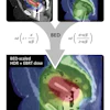


![A 53-year-old patient (patient number four) with a recurrent pituitary adenoma with extension of a cystic component of disease to the medial temporal lobe apparent on MRI (contoured in blue), and extension of disease to the left sphenoid bone and orbital apex apparent on [68Ga]Ga-DOTA-TATE (contoured in yellow).](https://img.auntminnie.com/mindful/smg/workspaces/default/uploads/2026/04/pituitary-tumor.QGsEnyB4bU.jpg?auto=format%2Ccompress&dpr=2&fit=crop&h=167&q=70&w=250)


