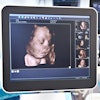
Three-dimensional ultrasound can be a valuable clinical tool, but its successful use requires awareness of the artifacts commonly encountered with the technique, said Dr. Dolores Pretorius of the 3D Ultrasound Imaging Group at the University of California, San Diego.
"Correct identification of artifacts is essential," she said. "Artifacts may mimic abnormal development, masses, or missing structures, and in OB (cases), I'm worried that babies are going to get terminated because someone sees something that they're sure is whatever they think it is."
Pretorius made her comments during a talk on artifacts, networking, and future perspectives on 3D ultrasound at the 2002 Leading Edge in Diagnostic Ultrasound meeting, held in Atlantic City.
The sources for 3D ultrasound artifacts can be B-mode artifacts (resolution, propagation, and attenuation matters), color or power Doppler artifacts (mainly from aliasing and gain), and 3D ultrasound-specific artifacts, Pretorius said.
Three-dimensional ultrasound-specific artifacts can be realized during the acquisition process from patient motion, such as respiration or fetal motion. Organ motion, such as vessel pulsations, cardiac motion, and peristalsis, can produce artifacts as well, she said.
Position-sensing errors can also have an impact. Metallic distortion can be produced by metal in the region of interest or in the area around your table if a magnetic sensor is being used to acquire the data, she said.
Improper calibration and operation will also produce poor data, Pretorius said. In addition, some 3D systems don't offer position-sensing capability, which can cause problems if caution is not exercised.
"Those (systems) can get into improper geometry if you're not very consistent with how you sweep," she said.
Rendering can also introduce artifacts, owing sometimes to too much or too little thresholding. Overthresholding can produce a "black eyes" artifact, for example.
The most common rendering artifacts users will see, however, are those created by region-of-interest boundaries, Pretorius said. If an ROI boundary is placed across the head, for example, it will appear as if the baby has a hole in the head.
The editing process can cause problems as well.
"When you edit 3D volumes and use those 'magic' scalpels, you can delete things that you shouldn't be deleting, and it'll look like there's an abnormality when it’s not," she said.
With that in mind, users need to confirm abnormalities identified on rendered images, either with multiplanar images or on repeat 2D scanning.
"You can't just (base your decision) on a new technology that's never been used before, and you just saw it for the first time, and (now) you're going to go for it," Pretorius said.
By Erik L. RidleyAuntMinnie.com staff writer
July 8, 2002
Related Reading
Adding 3D to power Doppler US improves prostate cancer detection, March 25, 2002
3D ultrasound enables precise diagnosis of PCO, March 18, 2002
Comparison between 2-D and 3D ultrasound measurements of nuchal translucency, January 4, 2002
3D ultrasound of fetal brain aids focused postnatal management, December 18, 2001
3D shoulder ultrasound flexes its muscle, June 4, 2001
Copyright © 2002 AuntMinnie.com


