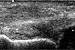
High-resolution ultrasound may be more accurate than initial clinical impression for evaluating focal lesions of the hand and wrist, according to research presented at the 2002 RSNA meeting in Chicago.
To determine the diagnostic accuracy of both techniques, a study team from the Mallinckrodt Institute of Radiology in St. Louis retrospectively reviewed the medical records of 77 consecutive patients between June 1996 and December 2001. The patients, identified from a computer search, had hand or wrist symptoms and received an ultrasound study followed by correlative surgery that identified a focal lesion, said Dr. Vikram Patel.
The diagnoses included:
- cystic lesion, including ganglion (59) and miscellaneous (3)
- solid lesion, including giant cell tumor (7), nerve tumor (3), hemangioma (2), and miscellaneous (3)
- focal synovitis (8)
- arterial thrombosis (1)
Ultrasound correctly diagnosed 65 of 77 lesions (84%), including 90% of cystic lesions, 67% of solid lesions, 89% of synovitis, and the one vascular lesion. Ultrasound correctly diagnosed solid lesions in all 15 cases, but gave the specific diagnosis in 10 of 15 cases, he said.
It did not detect the four ganglions and misdiagnosed two other cystic lesions. Of the five misdiagnosed ultrasound cases, the ultrasound study suggested a solid lesion, but the specific diagnosis was incorrect, Patel said.
In comparison, the initial clinical impression was concordant with pathologic findings in 48 of 77 cases (62%). In 38% of cases, there was no clinical impression given or there was discordance between the clinical diagnosis and the pathologic findings.
In seven cases, a solid tumor was misdiagnosed as a ganglion in the initial clinical impression. In three cases, a ganglion was misdiagnosed as a solid tumor. Solid, cystic, inflammatory, or vascular lesions were mistaken for each other in the remaining cases, according to Patel.
Overall, ultrasound was more accurate than clinical impression in evaluation of focal lesions of the hand or wrist. Moreover, the imaging modality can accurately determine synovitis cases, and yields specific diagnosis in most cases, especially if the lesion is cystic, he said.
"It also provided the correct diagnosis in 72% of cases where an initial clinical impression was not given or was discordant after pathology findings," Patel said. "This information allows the hand surgeon to more precisely discuss therapeutic options and outcomes with the patient."
By Erik L. RidleyAuntMinnie.com staff writer
February 28, 2003
Related Reading
Ultrasound detects occult scaphoid fractures, June 26, 2002
Electrodiagnostic testing best for diagnosing carpal tunnel syndrome, June 11, 2002
Copyright © 2003 AuntMinnie.com


