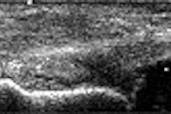
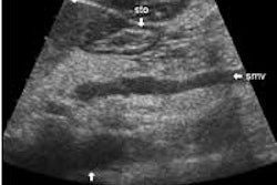
(Ultrasound Review) The sonographic features of Meckel’s diverticulum were described in the February issue of American Journal of Roentgenology. According to researchers at Hospital da Criança Conceição in Porto Alegre,Brazil, a tubular structure on sonography is suggestive of a Meckel's diverticulum in children presenting with rectal bleeding due to diverticulitis. They described the ultrasound, clinical, and pathologic findings in a retrospective study of ten children that had grayscale and color Doppler ultrasound.
Meckel’s diverticulum is a congenital intestinal abnormality, which usually is clinically challenging. Scintigraphy is useful in children with heavy rectal bleeding, but less sensitive in those with less intense bleeding. The authors reported "50% of children who are symptomatic present with an acute abdomen, and the diagnosis can be made only at surgery." Presenting symptoms were as follows: nine had abdominal pain, six clinically had acute appendicitis, and two had rectal bleeding.
Sonographic appearances are similar for acute appendicitis, intestinal duplication and inflamed Meckel’s diverticulum, but for the latter the mucosal layer is more irregular and 20% of patients had a cyst. Ultrasound was performed using a variety of linear and curved array transducers ranging in frequency from 7.5-3.5 MHz.
"In six patients, the inflamed Meckel's diverticulum presented as a fixed, noncompressible hypoechoic structure in the shape of a cul-de-sac in the right iliac fossa next to the anterior abdominal wall, with a diameter ranging from 0.8-1.2 cm," they reported.
One patient showed a complex mass thought to represent a perforated acute appendicitis. An oval cystic structure with typical but irregular gut wall was demonstrated in the right iliac fossa in two patients, and color Doppler showed increased vascularity and an anomalous artery. One patient with surgically proven Meckel's diverticulum was not detected sonographically. Ultrasound demonstrated distended loops of small bowel and increased peristaltic activity in this patient.
"We do not believe that sonography will supersede 99mTc pertechnetate scintigraphy because scintigraphy is a highly accurate tool to use in establishing the diagnosis of an inflamed Meckel's diverticulum," they reported.
However, they recommended that children with rectal bleeding and a negative scintigraphic proceed to ultrasound examination. The authors concluded "in patients in whom appendicitis is clinically suspected, the finding of a hypoechoic tubular structure in the iliac fossa on sonography is not a specific sign of appendicitis, and the possibility of an inflamed Meckel's diverticulum must be considered in the differential diagnosis."
Sonographic findings of Meckel’s diverticulitis in childrenBaldisserotto, M et al
Departmento de radiologia, Hospital da Criança Conceição--Ministério da Saúde, Porto Alegre, Brazil
AJR 2003 February; 180:425-428
By Ultrasound Review
March 27, 2003
Copyright © 2003 AuntMinnie.com


