Adenomyosis is frequently misdiagnosed as fibroids, resulting in inappropriate treatment and persistent symptoms, according to Dr. Edward Lyons, professor of radiology and obstetrics and gynecology at the University of Manitoba in Winnipeg, Canada.
"Until just a few years ago I was just as guilty of this mistake, but we went from virtually never making the diagnosis to making it six or seven times each and every single day. We are aware of the condition, we are aware of the specific sonographic findings, and the diagnosis is everywhere," Lyons said in an interview with AuntMinnie.com at the 2003 World Federation for Ultrasound in Medicine and Biology, and the American Institute of Ultrasound in Medicine meeting in Montreal.
Most clinicians and sonographers are unfamiliar with adenomyosis, which is defined as ectopic endometrial tissue similar to endometriosis, but located deep within the myometrium. Because of its location, it is sometimes referred to as endometriosis interna.
"I think that adenomyosis is terribly underdiagnosed," Lyons said. "The majority of people who come in for pelvic pain and menorrhagia have adenomyosis, but when people see diffusely enlarged uteri they write ‘myometrium has diffuse and homogenous changes likely consistent with multiple small fibroids.’ I’ve written that myself a million times and it was always wrong."
Lyons recommended using a transvaginal ultrasound probe for distinguishing adenomyosis from fibroids, as well as for assessing the patient’s pain.
"Using the probe as an extension of your examining finger, you will find areas, focal areas of uterine tenderness, that are usually associated with the abnormal areas that you see on the ultrasound," he said. "And fibroids are virtually never tender -- they are tender in two conditions, in pregnancy, and if they undergo infarction."
Whereas adenomyosis is the most common cause of pelvic pain, fibroids are more commonly the cause of abnormal bleeding. However, although about 80% of adenomyosis patients report pain, a full 20% do not, making other diagnostic tools important.
"One of the things I stress is to look at the entire package: look at the clinical, sonographic, and physical findings. With adenomyosis you have women who have usually had children, who have heavier than normal periods, often with clots, pain with their periods, and usually painful intercourse. The sonographic findings are this asymmetrically thickened endometrium, often areas of increased echogenicity and small cysts -- even 3 mm, subendometrial, myometrial, or intramural cysts."
This latter finding is often a source of confusion, Lyons said. "Many people see these small myometrial cysts and report them as ‘consistent with degeneration in a small fibroid.’ That is absolutely wrong, absolutely incorrect. These are distended endometrial glands and they are absolutely typical of adenomyosis."
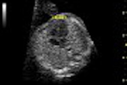
On ultrasound, adenomyosis appears as a diffuse, infiltrative process with central vessels, streaky shadows, and no calcification. In addition, cysts are often present.
Fibroids, in contrast, appear as well-defined masses and do not exhibit the myometrial inhomogeneity so often ascribed to them. They have a hypoechoic periphery due to the compressed myometrium. They can be hypoechoic, isoechoic, or hyperdense. They also have peripheral vessels, distal shadowing, and calcification.
Unlike adenomyosis, fibroids are better seen on transabdominal ultrasound than transvaginal, Lyons said. Making the correct differential diagnosis is important for treatment: Fibroid treatment, such as endometrial ablation, could be contraindicated in women with adenomyosis.
"We’ve seen several cases of women who end up with hematometras -- focused areas of increased bleeding, which get trapped into a partially obstructed portion of the endometrium after endometrial ablation and can cause increasing pain and tenderness," Lyons said.
Additionally, uterine artery embolization (UAE) will not alleviate adenomyosis because the latter is a diffuse infiltrative process with multiple vessels coming and going. In comparison, fibroids have a relatively small vascular pedicle. As soon as one artery is occluded, the fibroid is sensitive to ischemia and dies.
"(In adenomyosis), if you embolize the uterine artery, there are still many other vessels and there is no effect," Lyons explained.
Instead, adenomyosis is best treated medically with pain medication and high-dose birth control pills, such as Lupron or Danazol. Lyons said Danazol is less popular in the U.S. because of its reported side effects such as weight gain, hirsutism, and acne. But he suggested patients start on a low dose and use it sporadically to control their symptoms.
"Once their symptoms disappear, and that is usually very quickly, within one week, we find they can stop the Danazol for a while until their symptoms return -- so they’re actually only using it cyclically," he said.
By Kate JohnsonAuntMinnie.com contributing writer
July 18, 2003
Related Reading
Diffuse process on US related to extent of adenomyosis, September 4, 2002
Adenomyosis: Making the diagnosis with ultrasound, November 13, 2001
Copyright © 2003 AuntMinnie.com


