(Ultrasound Review) Color Doppler ultrasound provided additional essential information during biopsy of chest tumors, according to investigators from Atatürk University in Erzurum, Turkey.
The usual method for obtaining tumor cells in chest lesions is broncoscopy, however occasionally this can be difficult particularly in mediastinal and peripheral pulmonary lesions. An alternate diagnostic method is transthoracic needle aspiration (TTNA) using fluoroscopy, CT or ultrasound for guidance.
Dr. Metin Gorguner and colleagues prospectively studied 48 patients with mediastinal or peripheral pulmonary tumors. Their objective was to establish the diagnostic value of ultrasound guided TTNA of lung and mediastinal masses using color Doppler.
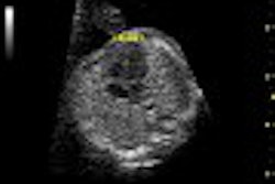
They discovered that incorporating color Doppler proved useful for two reasons. Firstly, it was used to demonstrate the vascular structures within the tumor in order to reduce the risk of penetrating large vessels during biopsy. Secondly, color Doppler was used to increase the conspicuity of the needle tip using the color twinkling sign. By moving the inner stylet of the needle, a strong Doppler frequency shift was created that resulted in color signals at the needle tip. This sign initially was described for renal calculi, and used to differentiate calcification from other echogenic structures within the kidney.
Comparing B-mode and color Doppler for detecting significant vessels, color Doppler showed vessels in 37 cases while B-mode alone showed vessels in 10 cases. Incorporating color Doppler provided a sensitivity of 90.0% and a specificity of 87.5% for obtaining a cytologic or microbiological diagnosis for TTNA.
Possible complications associated with this procedure include hemoptysis and pneumothorax, they reported. Other researchers have reported complications in up to 40% of TTNA cases without color Doppler, but there only were two partial pneumothoraxes in this study.
They recommended using small 21-gauge needles or less for this procedure. "In four cases a Tru-cut biopsy needle had to be used subsequently to obtain sufficient material for examination. When a Tru-cut biopsy needle has to be used, special care must be taken because of the risk of complications," they warned.
The authors concluded, "color Doppler sonography enables vascular structures within the tumor to be differentiated and provides real-time visualization of the needle tip, thus minimizing complications such as tissue hemorrhage and hemoptysis and increasing the quality of the material obtained." In their view, color Doppler ultrasound guidance of thoracic tumors reduces the risk of complication particularly for peripherally located tumors.
Color Doppler sonographically guided transthoracic needle aspiration of lung and mediastinal massesGorguner, Metin, et. al.
Departments of chest diseases and radiology, faculty of medicine, Atatürk University, Erzurum, Turkey.
J Ultrasound Med 2003 July; 22:703-708
By Ultrasound Review
August 26, 2003
Copyright © 2003 AuntMinnie.com


