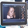
Three-dimensional ultrasound has potential as a valuable adjunct in pediatric and neonatal neurosonography, according to research published in European Radiology.
The technique "reduces imaging time, improves demonstration of complex anatomy and vasculature, and allows for evaluation of anatomy/pathology in any plane," wrote a research team led by Dr. Michael Riccabona from the University Hospital LKH Graz in Graz, Austria. "The 3-D ultrasound furthermore improves volume assessment (e.g. in hydrocephalus), (as well as) comparison with CT, MRI, and during follow-up, with a potentially improved standardization and documentation. Three-dimensional ultrasound additionally offers an ideal modality for training and education, as the brain and the neonatal spine can be virtually rescanned at the workstation" (European Radiology, September 2003, Vol. 9, pp. 2082-2093).
Riccabona and colleagues from the University of California, San Diego and the University Hospital LKH Graz discussed the potential of 3-D ultrasound from their experience with 150 patients using different 3-D techniques and three different 3-D ultrasound systems, comparing their experience with 2-D ultrasound studies, other imaging studies, and the literature.
Conventional two-dimensional ultrasound suffers from its operator-dependent nature, with a relatively high inter-/intra-observer variability that limits standardization and comparison during follow-up. In addition, it is difficult or impossible to acquire true axial planes and many oblique planes, the authors wrote.
"Critical sections such as the axial plane cannot be reliably evaluated," they stated. "This creates limitation during follow-up or comparing 2-D ultrasound to other modalities such as CT or MRI, and may obscure subtle changes in slowly evolving disease such as slowly growing hydrocephalus. Additionally, these 2-D ultrasound limitations degrade the diagnostic confidence in some conditions, as the problem is only partially addressed by using a transtemporal approach."
Transfontanellar 3-D ultrasound of neonatal brain
The benefits of 3-D ultrasound are evident, for example, with transfontanellar 3-D ultrasound of the neonatal brain. In the researchers’ experience, 3-D ultrasound was accurate, did not miss any essential diagnosis, and in individual cases, improved diagnostic confidence and/or helped with deciding on equivocal findings such as subtle subependymal hemorrhage, the authors said.
In addition, 3-D ultrasound added a number of other benefits, including demonstrating the often-complex anatomy of hydrocephalus and measuring ventricular volume, not achievable by 2-D ultrasound, they said. The addition of orthogonal and oblique planes gained with 3-D studies render the technique superior to 2-D exams in evaluating and demonstrating complex anatomic relations, as seen in some congenital brain malformation or post-hemorrhagic lesions, as well as in comparison with CT/MRI studies.
Evaluation of drain position in shunted hydrocephalus is improved with 3-D ultrasound, and its ability to provide an axial plane may make the modality superior in demonstrating and assessing extra-axial fluid collections (if accessible from original 2-D acquisition and included in 3-D volume), the researchers wrote.
Transfontanellar and transtemporal 3-D ultrasound in infants
Harmonic imaging may improve tissue differentiation on 2-D ultrasound, providing better data for 3-D reconstruction, according to the researchers.
If image acquisition is successful, 3-D ultrasound often provides better anatomic orientation in such conditions than 2-D ultrasound, particularly in situations where transfontanellar access is severely restricted or only transcranial imaging can be performed.
As transtemporal 3-D studies of cerebral vasculature improves visualization of vessel continuity and anatomy, particularly in tortuous vessels, it therefore improves visualization of vascular anatomy.
"This ability may support ultrasound in becoming a screening tool in an equivocal patient population, or may be used for follow-up of patients," the authors wrote.
Transcranial sonographic imaging of child’s brain
In this application, 3-D ultrasound reconstruction of segmented color data provides "superb visualization" of the major cerebral vessels, the authors stated. Three-dimensional ultrasound improves demonstration of the course of even a tortuous vessel, and allows for superior assessment of anatomical relationship and size.
These capabilities lead to improved depiction of an aneurysm or stenosis, and may be clinically useful for planning additional imaging, evaluating ICU patients, or following up patients with cerebral arteriovenous malformation.
Spinal sonography
Three-dimensional ultrasound may be beneficial in evaluating spine pathology in neonates and infants, provided a good-quality acquisition is obtainable, the authors said. High-resolution transducers are essential for high-quality 3-D reconstructions.
"Then 3-D ultrasound may offer not only a superior demonstration of the often-complex anatomic relationship, but additionally enables comprehensive visualization of skeletal structures using maximum intensity or ‘x-ray’-weighted transparent rendered views," they wrote.
The authors also acknowledged the drawbacks of 3-D ultrasound, including potential errors, limited spatial and gray-scale resolution, artifacts and misregistration, and technical limitations.
"Bearing these factors in mind, we propose a cerebral ultrasound protocol that may begin with an orienting 2-D ultrasound scan, followed by acquiring a few 3-D ultrasound data sets (in different projections), concluding the investigation with additional 2-D ultrasound of critical parenchymal areas (e.g. periventricular space) and/or (color Doppler/duplex Doppler sonography) evaluation of the cerebral vessels when indicated," they concluded. "With this approach, we believe 3-D ultrasound will become a valuable adjunct to neonatal and pediatric neurosonography."
By Erik L. RidleyAuntMinnie.com staff writer
November 4, 2003
Related Reading
No additional info offered by 3-D sonography of adnexal masses, June 19, 2003
3-D ultrasound offers reliable assessment of gallbladder, bile ducts, May 5, 2003
Power Doppler US spots tumor vascularity for prostate cancer staging, April 30, 2003
Contrast-enhanced 3-D US boosts mammography results, April 11, 2003
3-D power Doppler challenges standard aneurysm visualization, April 2, 2003
Copyright © 2003 AuntMinnie.com


