
ATLANTIC CITY - Although not yet approved by the Food and Drug Administration for use in radiology applications, ultrasound contrast media can significantly improve liver ultrasound, according to a presentation Tuesday at the Leading Edge in Diagnostic Ultrasound conference.
"With contrast agents, ultrasound has become competitive with CT and MRI in lesion detection, and may even be superior in lesion characterization and assessment of tumor viability following focal therapy," said Dr. Yuko Kono from the University of California, San Diego.
Contrast-enhanced ultrasound (CEUS) can dramatically change the role of ultrasound in liver tumor characterization, assessment of treatment response, evaluation of vascular problems, and diagnosis of solid organ injuries, she said.
Liver tumor characterization is the most important and widely used application of CEUS, she said. While some agents have been approved for radiology applications in Europe, Canada, and Asian countries, none have yet been cleared in the U.S.
For liver tumor detection, CEUS is an excellent tool for detecting metastasis. Even better performance is turned in with a liver-specific agent, Kono said.
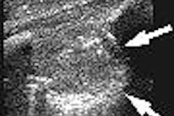
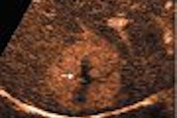
CEUS also shines in characterizing focal nodular hyperplasia (FNH). In fact, FNH is probably best characterized with CEUS, since the central feeding artery and spoke wheel vessels are better seen because of the high spatial and temporal resolution, Kono said.
Detecting hepatocellular carcinoma (HCC) is difficult, however, as not all HCC's are hypervascular.
"It's not a homogenous status in a very heterogenous liver, so it's not very simple," Kono said.
CEUS is also accurate for detecting residual tumor for assessing treatment response to radiofrequency ablation, and has been shown to be useful when performed interoperatively, she said.
Evaluating tumor viability
For assessing the results of transarterial chemoembolization (TACE), residual blood flow on CEUS after TACE is predictive of tumor outcome, Kono said. And it can be performed within days, as opposed to the delays needed for reliable detection using CT or MRI.
"We usually have to wait a month for CT," she said. "And to be reliable with MRI, which is more sensitive than CT, we have to wait three months."
UCSD recently studied the accuracy of post-TACE CEUS in 37 tumors. CEUS accurately detected tumor recurrence in all cases, with one false-positive, Kono said.
For evaluating liver trauma, CEUS delivers a performance comparable to enhanced CT, and can distinguish lacerations from contusions, she noted.
"But (the procedure) may be limited by patients' discomfort in this population," Kono said.
CEUS also can do a great job in diagnosing many vascular problems, when the lesion is deep or if there's not enough blood flow to produce a confident diagnosis, she said.
The modality is useful in applications such as evaluating the patency of transjugular intrahepatic portosystemic shunts, as well as vascular complications after liver transplantation.
Ultrasound contrast media will be critical in liver imaging, although it can't be used yet in the U.S. for radiology applications, Kono concluded.
"There are many applications in liver imaging," she said.
Kono also noted that power settings and frame rates are important to highlight regions with different blood flow and/or fractional blood volume.
By Erik L. Ridley
AuntMinnie.com staff writer
May 11, 2005
Related Reading
Contrast US pinpoints focal liver lesions, results in cost-savings, March 25, 2005
Breath-hold technique comparable to respiratory-triggered MR for liver imaging, March 16, 2005
Exam technique critical in liver ultrasound, October 22, 2004
Copyright © 2005 AuntMinnie.com








![Examples of ultrasound findings and techniques. (A) Images in a 39-year-old male patient with a mass in the left thigh. The mass is heterogeneous on the B-mode US image (compared with the patient in D) and showed increased microvascularity (superb microvascular imaging [SMI]) and shear-wave elastography (SWE) values. Undifferentiated pleomorphic sarcoma was diagnosed at biopsy (with pleomorphic rhabdomyosarcoma in surgical specimen). (B) Images in an 18-year-old male patient with a mass in the left leg. The mass is hypoechoic on the B-mode image, with no other findings suggestive of malignancy. The lesion is in contact with the cortex of the tibia, which is slightly irregular. CT revealed a doubtful anteromedial tibial erosion. The microvascular study demonstrated high vascularization, suggestive of malignancy. Periosteal Ewing sarcoma was diagnosed with both histologic and immunohistochemical confirmation. (C) Images in a 69-year-old female patient with a lump growing on the outside of the left leg. Multiple SWE examinations were performed (please note the high values obtained in the measurements, whereas the color map highlights the stiffness relative to adjacent tissues). SMI showed areas of increased vascularization to target for sampling. Undifferentiated spindle cell sarcoma was diagnosed at biopsy, with residual leiomyosarcoma in the surgical specimen after neoadjuvant therapy. (D) Images in a 56-year-old female patient with a mass in the right thigh. The mass is heterogeneous at both B-mode ultrasound (similar to patient A) and MRI (coronal T2-weighted spectral attenuated inversion recovery [SPAIR]; T1-weighted pre-contrast and postcontrast imaging), which even shows uptake after the administration of paramagnetic contrast material, which is traditionally suggestive of malignancy. Low values at SMI and elastography are suggestive of benignity. Spindle cell lipoma was diagnosed at biopsy, with atypical spindle cell lipomatous tumor in the surgical specimen.](https://img.auntminnie.com/mindful/smg/workspaces/default/uploads/2026/08/images-radiol250278fig2.APCFLSvX6p.jpg?auto=format%2Ccompress&dpr=2&fit=crop&h=167&q=70&w=250)
