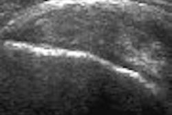
Contrast-enhanced sonography outperforms noncontrast-enhanced sonography in visualizing liver and spleen injuries, according to research published in the American Journal of Roentgenology.
To compare the injury detection rates and characterization of imaging findings of both techniques for confirmed solid organ injuries, Dr. John McGahan and colleagues from the University of California, Davis, School of Medicine performed a prospective study involving 22 injuries in 20 patients (AJR, September 2006, Vol. 187:3, pp. 658-666).
After hepatic, splenic, and renal injuries were identified on contrast-enhanced CT, both contrast-enhanced sonography and noncontrast-enhanced sonography (Sequoia, Siemens Medical Solutions, Malvern, PA; Definity, Bristol-Myers Squibb Medical Imaging, North Billerica, MA) were performed to identify the possible injury and analyze its appearance. The sonographic appearance of hepatic, splenic, and renal injuries were analyzed, and conspicuity was graded on a scale of 0 (nonvisualization) to 5 (high visualization).
Noncontrast-enhanced sonography showed 11 (50%) of the 22 injuries, while contrast-enhanced sonography revealed 20 (91%) of the 22 injuries. The only injury missed on contrast-enhanced sonography was a 2.5-cm laceration in the left lobe. However, during retrospective review of the video record, the laceration could be identified as a linear hypoechoic region, according to the researchers.
The average conspicuity grade for splenic injuries was 2.33 for contrast-enhanced sonography, compared with 0.67 for noncontrast-enhanced sonography. For liver injuries, contrast-enhanced sonography produced an average conspicuity grade of 2.2, compared with a 1.0 for noncontrast-enhanced sonography. Injuries that were missed resulted in a score of 0.
The researchers acknowledged several limitations in the study, including the use of reviewers who were aware that an injury had been identified on CT. Also, not all CT planes could be exactly reproduced by sonography because of the overlying ribs, and the series was limited in the number of injuries. In addition, there was no surgical correlation.
Contrast-enhanced sonography performed better than noncontrast-enhanced sonography for detecting solid organ injuries, the authors concluded. In addition, contrast-enhanced CT is the gold standard for evaluating blunt abdominal trauma patients, and remains the study of choice in patients who are hemodynamically stable.
Noncontrast-enhanced sonography continues to have an important role in triaging blunt abdominal trauma patients who are not hemodynamically stable and cannot undergo CT, according to the authors. As proposed by other researchers, contrast-enhanced sonography may also have a role in the initial evaluation of patients with blunt abdominal trauma.
"Certainly, contrast-enhanced sonography can be used in the follow-up of hospitalized patients with a known solid organ injury who are managed conservatively and who cannot be easily moved to the CT suite," the authors wrote. "Contrast-enhanced sonography could be used to help detect any changes in the injury and spare the patient further radiation exposure."
By Erik L. Ridley
AuntMinnie.com staff writer
October 5, 2006
Related Reading
Contrast-enhanced US picture shows signs of brightening, April 18, 2006
CEUS aids differentiation of small liver lesions, April 3, 2006
Detection of liver metastases with contrast-enhanced ultrasound, March 23, 2006
Contrast-enhanced US useful for differentiating focal liver lesions, January 19, 2006
Copyright © 2006 AuntMinnie.com


